Wikipedia:WikiProject Medicine/Cochrane/Cochrane Skin/Pictures of Skin Diseases
Welcome to the Cochrane Skin Pictures of Skin Diseases Project Page
Project Overview
[edit]Aim
[edit]To increase the number of photos of human skin diseases appearing in the Cochrane Library and Wikipedia.
Objective
[edit]To increase the number of photos of skin disease, including those of skin of different colours, in each Cochrane Skin Systematic Review and Wikipedia Skin disease article.
Approach
[edit]We will solicit skin disease pictures from Cochrane Skin consumers globally for addition to Cochrane Skin Systematic Reviews in the Cochrane Library and Wikipedia via the social media outreach. Pictures will need to be of acceptable quality as judged by our medical and technical experts and come with:
- a) appropriate permission to post the picture(s) and
- b) information that supports the diagnosis of the disease (such as a biopsy pathology report).
Progress Tracking:
[edit]Images Posted: Dermatologic disorders on Wikipedia
[edit]- Epidermoid cyst/Epidermal Inclusion Cyst
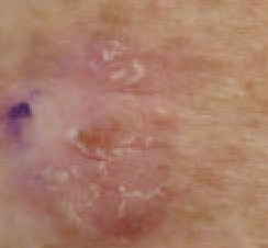
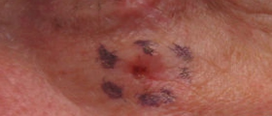
- Squamous cell skin cancer/Squamous Cell Carcinoma
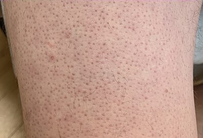
- Keratosis pilaris/Keratosis Pilaris
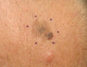
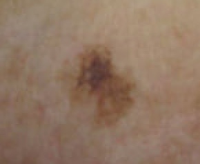
- Melanoma/Malignant Melanoma
Images Needed: Dermatologic disorders on Wikipedia
[edit]Here is a list of dermatologic disorders on Wikipedia that would benefit from additional clinical images.
- Amelanotic melanoma - a single image of a canine lesion
- Pemphigus vulgaris - single clinical image
- Bullous pemphigoid- single clinical image
- Urticarial vasculitis- no clinical images
- Chloracne- single image (not very helpful)
- Hidradenitis suppurativa- lacks images of each Hurley stage
- Dermatofibrosarcoma protuberans- no clinical images
- Pustulosis palmaris et plantaris- single image (not very helpful)
- Pityriasis rubra pilaris- single illustration, no clinical images
- Dermatosis papulosa nigra- no clinical images
Wikipedia's List of Skin Disorders
[edit]Cochrane Skin: Our Evidence
[edit]List of Cochrane Skin Reviews (Cochrane Library)
Progress
[edit]Images on Wikipedia Commons
[edit]-
Actinic keratosis, left upper paraspinal back (Original Post: Shared in Actinic keratosis)
-
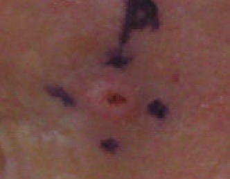
Basal Cell Carcinoma, left infraorbital cheek, marked for biopsy (Original Post: Shared in Basal-cell carcinoma)
-
Basal Cell Carcinoma, right cheek marked for biopsy (Original Post: Shared in Basal-cell carcinoma)
-
Basal Cell Carcinoma, ulcerated, nodular, on right lower cheek marked for biopsy (Original Post: Shared in Basal-cell carcinoma)
-
Epidermal Inclusion Cyst, nape of neck (Original Post: Shared in Epidermal inclusion cyst)
-
Inflamed epidermal inclusion cyst, nape of neck (Moved from Wikimedia Commons: Shared in Epidermal inclusion cyst)
-
Chronic folliculitis surrounding central sebaceous hyperplasia, right mid chest (Original Post: Shared in Folliculitis)
-
Keratosis Pilaris on Lower Extremity (Original Post: Shared in Keratosis pilaris)
-
Keratosis Pilaris on Back of Upper Arm (Moved from Wikimedia Commons: Shared in Keratosis pilaris)
-
Keratosis pilaris arm (Moved from Wikimedia Commons: Shared in Keratosis pilaris)
-
Lentigo Maligna Melanoma, Left Central Malar Cheek marked for biopsy (Original Post: Shared in Lentigo maligna) (Original Post: Shared in Lentigo maligna melanoma)
-
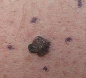
Malignant Melanoma, right posterior thigh (Original Post: Shared in Melanoma)
-
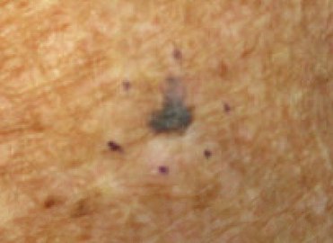
Melanoma in situ, vertex scalp marked for biopsy (Original Post: Shared in Melanoma)
-
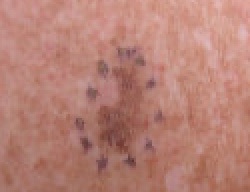
Malignant Melanoma in situ, evolving, right clavicle marked for biopsy (Original Post: Shared in Melanoma)
-
Malignant Melanoma, vertex scalp marked for biopsy (Original Post: Shared in Melanoma)
-
Malignant Melanoma, right medial thigh marked for biopsy (Original Post: Shared in Melanoma)
-
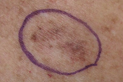
Malignant Melanoma, right posterior shoulder circled for biopsy (Original Post: Shared in Melanoma)
-
Malignant Melanoma, left forearm marked for biopsy (Original Post: Shared in Melanoma)
-
Malignant Melanoma left forearm post excision with purse string closure (Original Post: Shared in Melanoma)
-
Melanoma in situ, right forehead marked for biopsy (Original Post: Shared in Melanoma)
-
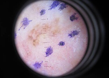
Melanoma in situ, dermatoscope image, right forehead marked for biopsy (Original Post: Shared in Melanoma)
-
Malignant Melanoma in situ, evolving, medial right temple with adjacent sebaceous hyperplasia, lateral (Original Post: Shared in Melanoma)
-
Malignant Melanoma in situ, left anterior shoulder marked for biopsy (Original Post: Shared in Melanoma)
-
Malignant Melanoma in situ, right anterior shoulder marked for biopsy (Original Post: Shared in Melanoma)
-
Malignant Melanoma in situ, left upper inner arm (Original Post: Shared in Melanoma)
-
Malignant Melanoma in situ, right upper medial back, marked for biopsy (Original Post: Shared in Melanoma)
-
Malignant Melanoma in situ marked for biopsy, left forearm (Original Post: Shared in Melanoma)
-
Malignant Melanoma, mid frontal scalp (Original Post: Shared in Melanoma)
-
Malignant melanoma, left mid back marked for biopsy (Original Post: Shared in Melanoma)
-
Malignant melanoma, left mid back marked for biopsy, through dermatoscope (Original Post: Shared in Melanoma)
-
Compound nevus, left buttock (Original Post: Shared in Nevus)
-
Neurofibroma marked for biopsy, right upper back (Original Post: Shared in Neurofibroma)
-
Sebaceous hyperplasia, lateral right temple marked for biopsy with adjacent malignant melanoma in situ, evolving, medial right temple (Original Post: Shared in Sebaceous hyperplasia)
-
Sebaceous hyperplasia with surrounding chronic folliculitis, right mid chest (Original Post: Shared in Sebaceous hyperplasia)
-
Squamous Cell Carcinoma, right upper cheek; Lesion outlined in blue marker with a dashed line prior to biopsy (Original Post: Shared in Squamous Cell Carcinoma)
-
Squamous Cell Carcinoma, well differentiated, left upper paraspinal back marked for biopsy with adjacent actinic keratosis (Original Post: Shared in Squamous Cell Carcinoma)
-
Squamous Cell Carcinoma, left lateral canthus marked for biopsy (Original Post: Shared in Squamous Cell Carcinoma)
-
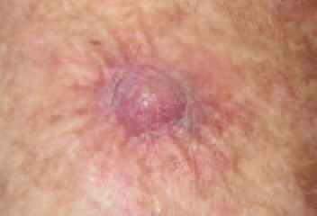
Squamous Cell Carcinoma, left ventral forearm (Original Post: Shared in Squamous Cell Carcinoma)
-
Tinea Corporis, right buttock (Original Post: Shared in Tinea corporis)
Resources
[edit]How to add images to Wikipedia Commons
[edit]1. Register for a Wikipedia Account:
2. Ensure your images are suitable for Wikipedia Commons. See Commons:Contributing your own work for more information
3. Log into Wikipedia Commons using your Wikipedia Account and upload your images:
Tips for preparing your images
[edit]- Ensure that your photo does not contain locator information. You can do this by either turning off the locator on your phone or by taking a screenshot of your image on your computer, saving it as a new jpeg, and uploading the new version only to commons.
- Still shots are required, jpeg format seems to work well.
- Include a description of your photograph. See examples in the gallery above. The more information you provide, the easier it will be to find an appropriate place to share your photo in a Wikipedia article.
- Be sure to speak with your institution in order to use the appropriate permission form to obtain written consent.
- The person should not be identifiable from the photograph so please zoom in or crop prior to uploading on Commons.
- Please add your new photos to our gallery, above.
- Wikipedia Commons essay with information on sharing images of patients: https://commons.wikimedia.org/wiki/Commons:Patient_images
Where can an image be shared on Wikipedia?
[edit]- Wikipedia's Medical Manual of Style Guideline gives suggestions for sharing images in a gallery: WP:MEDMOS#Images
- Images may also be appropriate in the Medical Info Box at the top of the article. https://wiki.riteme.site/wiki/Help:Infobox

