Wikipedia:Picture peer review/Human embryogenesis
Appearance


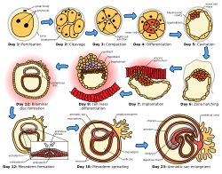
Highly encyclopedic and illustrative, clearly showing the important stages in the first three weeks of formation of the human embryo. However, it probably needs converting to svg before submitting to FPC, and I don't have a clue how to do that...
- Articles this image appears in
- Human embryogenesis, Prenatal development + 8 others
- Creator
- Jrockley
- Suggested by
- Anxietycello (talk) 13:31, 30 May 2010 (UTC)
- Comments
- You can request the image be converted to .svg over at Wikipedia:Graphic Lab. Also, the image needs references. See FPC criterion 6: "Is accurate. It is supported by facts in the article or references cited on the image page, or is from a source noted for its accuracy." Finally, I think it might flow better if it went back and forth rather than resetting to the beginning of the line, so that 5 would be right below 4. That way it's easier to compare stages. Others might disagree there, though. Makeemlighter (talk) 22:56, 30 May 2010 (UTC)
- I've just added references to the image page, and put in a conversion request at the graphics lab. I might try creating an alt image as you described tomorrow. Thanks for the advice! Anxietycello (talk) 04:15, 31 May 2010 (UTC)
- I would suggest adding an approximate scale to each image too... - Zephyris Talk 15:32, 5 July 2010 (UTC)
- A second little point, from day 12 onwards it is not clear which side of the uterine epithelium the embryo is on... Which is it? - Zephyris Talk 15:37, 5 July 2010 (UTC)
- Vector version now available. - Zephyris Talk 20:46, 5 July 2010 (UTC)
- Seconder
- Post-vectorisation I would say this image has a good chance at featured picture status. It has huge EV, is widely used across many major articles and is of a quality comparable to other featured svg diagrams. Furthermore I am able to actively improve it if there are any concerns which need addressing. - Zephyris Talk 19:44, 7 July 2010 (UTC)
- Brilliant work! Thankyou very much for taking the time to do this. Only a few small niggling issues - a typo on day six (should read zona instead of zoa); the captions on day 12 and 23 are bold unnecessarily, and on day 23, could you make it clearer that the yolk sac is a single body (it currently appears too tightly pinched at its emergence form the embryo). Also, I know it may not be clear, but I intended the orange colouring to be used only for the 'inner cell mass'; once it splits into the endoderm and ectoderm, the cell mass was to be represented as pink and brown (ie skin and digestive lining), and so there should be no orange cells after day 7. Thankyou again for your excellent work, I'll nominate it for FP now. Anxietycello (talk) 12:57, 15 July 2010 (UTC)
- All those points should be fixed now... - Zephyris Talk 19:15, 15 July 2010 (UTC)
- Conclusion
Nominated at FPC here. Makeemlighter (talk) 03:46, 19 July 2010 (UTC)
