User:NadiaMichelleKhan/sandbox
| Inferior gluteal nerve | |
|---|---|
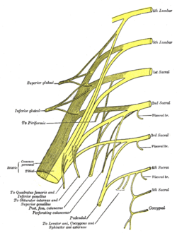
 Plan of sacral and pudendal plexuses. (Inferior gluteal nerve labeled at middle left.) | |
 The gluteus medius and nearby muscles | |
| Details | |
| From | sacral plexus (L5–S2) |
| Innervates | gluteus maximus |
| Identifiers | |
| Latin | nervus gluteus inferior |
| Anatomical terms of neuroanatomy | |
| This user page or section is in a state of significant expansion or restructuring. You are welcome to assist in its construction by editing it as well. If this user page has not been edited in several days, please remove this template. If you are the editor who added this template and you are actively editing, please be sure to replace this template with {{in use}} during the active editing session. Click on the link for template parameters to use.
This page was last edited by Avicenno (talk | contribs) 6 years ago. (Update timer) |
The inferior gluteal nerve is the main motor neuron that innervates the gluteus maximus muscle. It is responsible for the movement of the gluteus maximus in activities requiring the hip to extend the thigh, such as climbing stairs. Injury to this nerve is rare but often occurs as a complication of posterior approach to the hip during hip replacement. When damaged, one would develop gluteus maximus lurch, which is a gait abnormality which casues the individual to 'lurch' backwards to compensate lack in hip extension.
Anatomy
[edit]The largest muscle of the posterior hip, gluteus maximus, is innervated by the inferior gluteal nerve. (realtionship)It branches out which enter the deep surface of the gluteus maximus, the principal extensor of the thigh, and supply it. .
Origin of Nerve
[edit]The muscle is supplied by the inferior gluteal nerve which arises from the dorsal branches of the ventral rami of the fifth lumbar and the first and second sacral nerves. (2006 ling)
The inferior gluteal nerve originates from sacral plexus originates from the dorsal L5, S1, and S2 rami. -carries fiber from L5,S1, and S2, The inferior gluteal nerve arires from the dorasl diviisoins of the fifth lumbar and the first and sexond ventral rami. (new 2009 paper not citesd yet)
Arises from the ventral divisions of L5, S1 and S2
It arises from the posterior divisions of the ventral rami of L5 through S2
The lumbosacral_trunk , which is made up of L5 and a small branch of L4, effectively connects the lumbar and sacral plexuses.(mr) The lower branches of the L4 and the L5 nerves enter the sacral plexus
The sacral plexus is formed by the lumbosacral tmnk, the first to third sacral ventral rami, and part of the fourth, the remainder of the last joining the coccygeal plexus.^ The sacral plexus (Fig. 3) is formed in the pelvis in front of the piriformis muscle.
The sacral plexus is formed anterior to the piriformis muscle and gives rise to the sciatic nerve, the superior and inferior gluteal nerves, and the pudendal and posterior femoral cutaneous nerves.
However, most of the sacral plexus nerves are scarcely recognizable, because they leave the pelvis through the greater sciatic foramen. From the pelvis, the anterior primary branches of the nerves entering the plexus (the first sacral nerve being a particularly large one) and a mass of nerves on the piriformis can be recognized.'"
Course of Nerve
[edit]Leaves the pelvis through the greater sciatic foramen Runs underneath the piriformis Divides into muscular branches to supply the gluteus maximus It leaves the pelvis through the greater sciatic foramen below the pyriformis muscle.
traverses the greater sciatic foramen just inferior to the piriformis muscle
It leaves the pelvis through the greater sciatic notch below the piriformis muscle and divides into branches that pass posteriorly into the deep surface of the gluteus mazimus muscle.(new 2009)
Exits the pelvis through the greater sciatic foramen, superficial to the sciatic nerve. It has been described as having multiple branches with subsequent innervation of the overlying gluteus maximus (relationship)
The inferior gluteal nerve reliably emerged inferior to the piriformis muscle in all our specimens. The branching characteristics of the nerve fell into two broad categories. Of the 15 examined hips, nine demonstrated a short stalk which came under the piriformis and then gave rise to all of the terminal branches of the nerve which spanned the muscle of the gluteus maximus (Fig. 1). The number of branches from the stalk ranged from four to six. In the other six specimens, at least a partial split of the stalk occurred proximal to the coverage of the piriformis. In these specimens there were two to three divisions of the inferior gluteal nerve under the piriformis that would further divide close to the insertion of the nerve into the actual muscle belly (relationship)
The nerve was always seen close to and medial to the sciatic nerve when it left the sacral plexus inferior to the piriformis. In all specimens, the nerve entered the deep surface of gluteus maximus approximately 5 cm from the tip of the greater trochanter of the femur and entered the deep surface of gluteus maximus over the inferior one-third of the muscle belly. (2006 ling)
The inferior gluteal nerve is accompanied by the inferior gluteal artery, a branch of the anterior trunk of the internal iliac artery. (ling 2006)
-However The relationship between the inferior gluteal nerve and artery was found to be unpredictable in our study No consistent relationship between the inferior gluteal artery and the inferior gluteal nerve was observed in our current study
The proximal stalk or the proximal divisions of the inferior gluteal nerve, after they were distal to the piriformis, could be reliably located using the external anatomic landmarks shown in Fig. 3; the cephalo-caudal level was represented by a line connecting the most prominent lateral borders of the greater trochanters of both femurs. The medial-lateral level was defined by a line perpendicular to the cephalo-caudal level and centered at the ischial tuberosity. The depth was found to be at the level of the posterior border of the proximal femur. These landmarks reliably represented the targeted area—common beginnings of the innervation to the gluteus maximus muscle—in all 15 examined hips. The resultant target was also in a location that was isolated from the sciatic nerve. or this paragraph?
We did, however, observe a relationship between the common stalk of the inferior gluteal nerve and external anatomic landmarks. It is our current opinion based on these observations that the targeted region should be aimed inferior to the most prominent aspect of the greater trochanter, and medial to the landmark of the ischial tuberosity, at the depth of the posterior border of the proximal femur. Triangulating using these three coordinates, one can reliably reach the source of the inferior gluteal nerve. This will result in maximal stimulation of the gluteus maximus musculature when using electrical stimulation for the purpose of prevention of pressure ulcers.
The inferior gluteal nerve originates in the sacral plexus. It arises from the posterior divisions of ventral rami of fifth lumbar and first and second sacral nerves. It leaves the pelvis through the greater sciatic foramen, medial to the sciatic nerve. The inferior gluteal nerve courses below the piriformis muscle, while the superior gluteal nerve courses above the piriformis muscle. ,
The sciatic nerve (L4 to S3), the largest nerve of the body, immediately leaves the pelvis through the greater sciatic foramen, below the piriformis. The superior gluteal nerve passes backward through the greater sciatic foramen, above the piriformis: the inferior gluteal nerve also passes backward through the greater sciatic foramen but below the piriformis." -(surgery)
We noted that the inferior gluteal nerve entered the deep surface of gluteus maximus very inferiorly.
It lies medial to the sciatic nerve and exits the pelvis through the greater sciatic foramen, inferior to the piriformis muscle. At the lower border of the piriformis muscle, the nerve turns backward and divides into upward and downward diverging branches, which enter the gluteus maximus. The nerve may also send a branch to the posterior femoral cutaneous nerve. (MR)
Nerve Function
[edit]The major function of the gluteus maximus is to extend the flexed thigh and bring it inline with the trunk. It may prevent the forward momentum of the trunk from producing flexion at the supporting hip during bipedal gait. It is intermittently active in the walking cycle and in climbing stairs and continuously active in strong lateral rotation and abduction of the thigh and also stabilizes the femur on the tibia when the knee extensors are relaxed [14] [1] previously. In addition to this, the gluteus maximus has an important role during some activities like running or standing up.
gluteus maximus = strong hip extensor prevents jackknifing at the hip on heel strike hip stablizer
-gluteus maximus = major hip extensor and stabilizer of the trunk
-prevents trunk from falling forward
- gluteus maximus contracts at heel-strike, slowing forward motion of trunk by arresting flexion of the hip and initiating extension;
- extends thigh at the hip, assists in laterally rotating the thigh;
The gluteus maximus, a large muscle with numerous attachments, is a powerful extensor of the thigh or of the trunk lower limbs are in a fixed position. Surprisingly, however, it is not important posturally, is relaxed when one is standing, and is little used in walking. It is employed in running, climbing, and rising from a sitting or stooped position. It also controls flexion at the hip upon sitting down (paradoxical action).
Nerve Injury
[edit]The patients demand to preserve such activities after hip arthroplasty, thus, it is important to preserve these functions. (2009 course)
Due to Hip Replacement
[edit]Failure of these functions due to denervation of gluteus maximus would result in gait abnormalities.
Inferior gluteal entrapment neuropathy is rarely reported but is recognized as a complication of the posterior approach to hip arthroplasty. Injuries to the peripheral nerves occur in 0.5% to 8% of patients undergoing total hip displacement,
The posterior approach has been assessed most widely and is perhaps the most frequently used, but it is also the one most likely to be associated with damage to the inferior gluteal nerve since this structure is not usually seen. Direct abnormalities of the nerve may be difficult to detect due to the small size of the nerve, although signal intensity alterations in the gluteus maximus may be encountered (mr)
Diagnostic imaging of peripheral nerves about the hip is a challenging task due to the complex regional anatomy, the small size and intricate course of many nerves as well as the variety of clinical situations leading to local disturbances in the nerve function the posisition of the inferiror gluteal nerve makies it vulnerable to iagtrogenic injury durong posterior and postlatertl approaches to the hip
It is subject to injury by compression and ischemia in sedentary individuals, resulting in difficulty in rising from a sitting position and difficulty climbing stairs.
The incidence of damage to the inferior gluteal nerve after replacement of the hip is still uncertain.1 Peripheral nerve injury may occur during operations on the hip as a result of operative trauma associated with stretching and retraction of the nerve,
Few studies have focused on damage to the inferior gluteal nerve during hip replacement.
In ten other patients who had a posterior approach, nine had abnormal electromyographic findings in inferior gluteal innervated muscles and eight of the ten also had abnormalities in superior gluteal innervated muscles. They suggested that abnormalities of gait after the operation may be due to injury to these nerves. In another study, Perron et al9 concluded that the reduction in walking speed and persistently abnormal gait, sometimes seen in patients one year after THR, were associated with a decrease in the extensor moment with a resultant decrease in the range of extension of the hip and a reduction in the abductor moment.
When a muscle-splitting incision is made across gluteus maximus as part of the classical posterior approach11 and the muscle parted by hand-held or self-retaining retractors, the likelihood of damage to the inferior gluteal nerve is high The nerve enters the deep surface of the muscle and is not easily visualised and differentiated from other structures running with it, such as the blood vessels. Parting the muscle damages the nerve further by stretching or even rupturing its branches which run superiorly on its deep surface.
Entrapment neuropathy is an underrecognized cause of pain and functional impairment caused by acute or chronic injury to peripheral nerves.
Although nerves may be injured anywhere along their course, they are more prone to compression, entrapment, or stretching as they traverse anatomically vulnerable regions, such as superficial or geographically constrained spaces. Subclinical electromyographic abnormalities of both the superior and inferior gluteal nerves have been described in up to 77% of patients after total hip replacement, regardless of the surgical approach
The posterior approach is the most common and practical of those used to expose the hip joint. The posterior approaches allow excellent visualization of the femoral shaft, thus are popular for revision joint replacement surgery in cases in which the femoral component needs to be replaced [2, 4]. The likelihood of damage to the IGN is reported to be high when a muscle-splitting incision is made across the gluteus maximus as a part of the classical posterior approach to the hip.
This may cause selective denervation of the gluteus maximus since the IGN courses along the deep surface of the muscle and is not easily visualized and differentiated from other structures running with it, such as blood vessels
Gluteus Maximus Lurch
[edit]Symptom
Injury to this nerve leads to a gluteus maximus lurch when gluteus maximus is weak/injured, trunk extends (lean back) on heel-strike on weakened side compensates for weakness of hip extension
-ign = loss of extension at hip, buttock wasting
- gluteus maximus gait pattern: (see: gluteus maximus) - begins to contract at moment of heel-strike, slowing forward motion of trunk by arresting flexion of hip & initiating extension; - when gluteus maximus is weak, trunk lurches backward (gluteus maximus lurch) at heel-strike on weakened side to interrupt forward motion of the trunk;
Treatment & Rehabilitation of Fractures
Gluteus maximus lurch
-difficulty preventeding the flexion of the trunk heel strike -may use trunk entension before heel strike to matain balance causing a backwards lurch
gluteus maximus = strong hip extensor
prevents jackknifing at the hip on heel strike
hip stablizer
Gluteus Maximus Lurch (Hip Extensor) The trunk lurches back on the stance phase side hyperextending.
Gait Analysis In The Science Of Rehabilitation
-in weaknes of glut max, -indivdual will throw him backward with a 'lurch' using adominal andoaraspinal muscle activation just after heel strike on affected side -backwards trunk lurch presists through out the stance to maintain the gravintaional force line behind the hip axis locking the hip into extension -there is an apparent forward protursion of the affected hip due to exgrated hip mation and the person may also hold the sholders backward to keep yth center of grapgty behind the goint. Yhe hamstring muscles osten compentsate for the glut maz weakness resulting in a near normal gait pattern but most often theses muschesla re affexted to gether.
References
[edit]- ^ Petchprapa, C. N., et al. "Mr Imaging of Entrapment Neuropathies of the Lower Extremity Part 1. The Pelvis and Hip." Radiographics 30.4 (2010): 983-1000. Print.
- ^ Mondelli, M., et al. "Mononeuropathies of Inferior and Superior Gluteal Nerves Due to Hypertrophy of Piriformis Muscle in a Basketball Player." Muscle & Nerve 38.6 (2008): 1660-62. Print.
- ^ Dejong, P. J., and T. W. Vanweerden. "Inferior and Superior Gluteal Nerve Paresis and Femur Neck Fracture after Spondylolisthesis and Lysis - a Case-Report." Journal of Neurology 230.4 (1983): 267-70. Print.
- ^ Skalak, A. F., et al. "Relationship of Inferior Gluteal Nerves and Vessels: Target for Application of Stimulation Devices for the Prevention of Pressure Ulcers in Spinal Cord Injury." Surgical and Radiologic Anatomy 30.1 (2008): 41-45. Print.
- ^ Ling, Z. X., and V. P. Kumar. "The Course of the Inferior Gluteal Nerve in the Posterior Approach to the Hip." Journal of Bone and Joint Surgery-British Volume 88B.12 (2006): 1580-83. Print.
- ^ Tagliafico Alberto, et al. "Imaging Of Neuropathies About The Hip." European Journal Of Radiology (n.d.): ScienceDirect. Web. 16 Nov. 2012
- ^ Apaydin, N., et al. "The Course of the Inferior Gluteal Nerve and Surgical Landmarks for Its Localization During Posterior Approaches to Hip." Surgical and Radiologic Anatomy 31.6 (2009): 415-18. Print.
- ^ Mirilas, P., and J. E. Skandalakis. "Surgical Anatomy of the Retroperitoneal Spaces, Part Iv: Retroperitoneal Nerves." American Surgeon 76.3 (2010): 253-62. Print.
- ^ Hoppenfeld, Stanley (2000). Treatment and rehabilitation of fractures. Philadelphia [u.a.]: Lippincott Williams & Wilkins. pp. 39, 259, 277. ISBN 0781721970.
See also
[edit]External links
[edit]- Inferior gluteal nerve at the Duke University Health System's Orthopedics program
- Gluteus Maximus Lurch / Inferior Gluteal Nerve This video is a demonstration of gluteus maximus lurch caused by damage to the inferior gluteal nerve.
![]() This article incorporates text in the public domain from the 20th edition of Gray's Anatomy (1918)
This article incorporates text in the public domain from the 20th edition of Gray's Anatomy (1918)

