User:Gensurg22/sandbox
Current Articles
[edit] | This is a user sandbox of Gensurg22. You can use it for testing or practicing edits. This is not the sandbox where you should draft your assigned article for a dashboard.wikiedu.org course. To find the right sandbox for your assignment, visit your Dashboard course page and follow the Sandbox Draft link for your assigned article in the My Articles section. |
Ref_List
[edit]My Images for Donation
[edit]Notes and Drafting
[edit]Hereditary spherocytosis [DRAFTING]
[edit]From Wikipedia, the free encyclopedia Jump to navigation Jump to search
This article is about aspects of spherocytosis specific to the hereditary form of the disorder. For details that apply generally to this variant as well as others, see Spherocytosis.
| Hereditary spherocytosis | |
|---|---|
| Other names | Minkowski–Chauffard syndrome |
| Peripheral blood smear from patient with hereditary spherocytosis | |
| Specialty | Hematology |
Hereditary spherocytosis (HS) is a congenital hemolytic disorder, wherein a genetic mutation coding for a structural membrane protein phenotype leads to a spherical shaping of erythrocytic cellular morphology. As erythrocytes are sphere-shaped (spherocytosis), rather than the normal biconcave disk-shaped, their morphology interferes with these cells' abilities to be flexible during circulation throughout the entirety of the body - arteries, arterioles, capillaries, venules, veins, and organs. This difference in shape also makes the red blood cells more prone to rupture under osmotic and/or mechanical stress. Cells with these dysfunctional proteins are degraded in the spleen, which leads to a shortage of erythrocytes resulting in hemolytic anemia.
HS was first described in 1871, and is the most common cause of inherited hemolysis in populations of northern European descent, with an incidence of 1 in 5000 births. The clinical severity of HS varies from mild (symptom-free carrier), to moderate (anemic, jaundiced, and with splenomegaly), to severe (hemolytic crisis, in-utero hydrops fetalis), because HS is caused by genetic mutations in a multitude of structural membrane proteins and exhibits incomplete penetrance in its expression.
Early symptoms include anemia, jaundice, splenomegaly, and fatigue. Acute cases can threaten to cause hypoxia secondary to anemia and acute kernicterus through high blood levels of bilirubin, particularly in newborns. Most cases can be detected soon after birth. Testing for HS is available for the children of affected adults. Occasionally, the disease will go unnoticed until the child is about 4 or 5 years of age. A person may also be a carrier of the disease and show no signs or symptoms of the disease. Late complications may result in the development of pigmented gallstones, which is secondary to the detritus of the broken-down blood cells (unconjugated or indirect bilirubin) accumulating within the gallbladder. Also, patients who are heterozygous for a hemochromatosis gene may exhibit iron overload, despite the hemochromatosis genes being recessive.In chronic patients, an infection or other illness can cause an increase in the destruction of red blood cells, resulting in the appearance of acute symptoms, a hemolytic crisis. On a blood smear, Howell-Jolly bodies may be seen within red blood cells. Primary treatment for patients with symptomatic HS has been total splenectomy, which eliminates the hemolytic process, allowing normal hemoglobin, reticulocyte and bilirubin levels. The resultant asplenic patient is susceptible to encapsulated bacterial infection, and prevented with vaccination. If other symptoms, such as abdominal pain persist, the removal of the gallbladder may be warranted for symptomatic cholelithiasis.
Contents
[edit]- 1 Epidemiology
- 2 Etiology
- 3 Pathophysiology
- 4 Clinical Presentation
- 5 Diagnostics
- 6 Treatment
- 7 Complications
- 8 Research
- 9 See also
- 10 References
- 11 External links
Epidemiology
[edit]Hereditary spherocytosis is the heritable hemolytic disorder, affecting 1 in 2,000 people of Northern European ancestry. According to Harrison's Principles of Internal Medicine, the frequency is at least 1 in 5,000 within the United States of America. While HS is most commonly (though not exclusively) found in Northern European and Japanese families, an estimated 25% of cases are due to spontaneous mutations.
Etiology
[edit]Hereditary spherocytosis is an erythrocytic disorder of that affects the following red cell membrane proteins in a congenital fashion:
- Spectrin (alpha and beta)
- Ankyrin
- Band-3 Protein
- Protein-4.2
- Lesser proteins of significance
Hereditary spherocytosis can be an autosomal recessive or autosomal dominant trait. The autosomal recessive inheritance pattern accounts for close to 25% of the clinical cases. The autosomal dominant inheritance patter accounts for over 75% of the clinical cases. Many positive individuals will not present clinically, thus the etiologic data may be artificially skewed towards the more prominent dominant forms. These dominant forms tend to leave a family history that yields generational splenectomies and black gallstones cholelithiasis. Lastly, an estimated 25% of cases are due to spontaneous mutations.
Pathophysiology
[edit]Causative Mutations and Phenotypic Expressions
[edit]Hereditary spherocytosis is caused by a variety of molecular defects in the genes that code for the red blood cell proteins spectrin (alpha and beta), ankyrin, band 3 protein, protein 4.2, and other red blood cell membrane proteins:
| Hereditary Spherocytosis Type | Genotypic Etiology | Phenotypic Expression | ||
|---|---|---|---|---|
| OMIM* | Gene | Locus | Erythrocyte Membrane Protein Affected | |
| HS-1 | 182900 | ANK1 | 8p11.2 | Ankyrin |
| HS-2 | 182870 | SPTB | 14q22-q23 | Spectrin (Beta)* |
| HS-3 | 270970 | SPTA | 1q21 | Spectrin (Alpha-1)* |
| HS-4 | 612653 | SLC4A1 | 17q21-q22 | Band-3 Protein |
| HS-5 | 612690 | EPB42 | 15q15 | Protein-4.2 |
*Online Mendelian Inheritance in Man (OMIM). The Alpha-1 refers the Alpha-1 Subunit of the Spectrin protein. The Beta refers the Beta Subunit of the Spectrin protein. [TABLE FIXED]
Pathophysiology of Mutated Erythrocytic Membrane Proteins
[edit]These proteins are necessary to maintain the normal shape of a red blood cell, which is a biconcave disk. The integrating protein that is most commonly defective is spectrin which is responsible for incorporation and binding of spectrin, thus in its dysfunction cytoskeletal instabilities ensue.[citation needed]
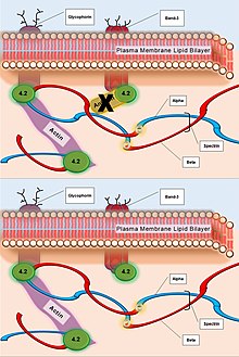
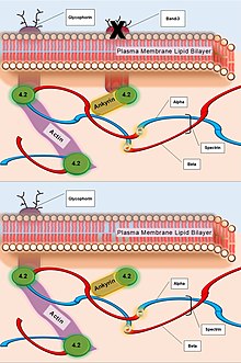
The primary defect in hereditary spherocytosis is a deficiency of membrane surface area. Decreased surface area may be produced by two different mechanisms: 1) Defects of spectrin, ankyrin (most commonly), or protein 4.2 lead to reduced density of the membrane skeleton, destabilizing the overlying lipid bilayer and releasing band 3-containing microvesicles. 2) Defects of band 3 lead to band 3 deficiency and loss of its lipid-stabilizing effect. This results in the loss of band 3-free microvesicles. Both pathways result in membrane loss, decreased surface area, and formation of spherocytes with decreased deformability.[citation needed]
[BUILD TABLE ON PAGE LIVE]
Cardiovascular and Organ Sequelae
[edit]As the spleen normally targets abnormally shaped red cells (which are typically older), it also destroys spherocytes. In the spleen, the passage from the cords of Billroth into the sinusoids may be seen as a bottleneck, where red blood cells need to be flexible in order to pass through. In hereditary spherocytosis, red blood cells fail to pass through and get phagocytosed, causing extravascular hemolysis.
Clinical Presentation
[edit]Complications
[edit]- Hemolytic crisis, with more pronounced jaundice due to accelerated hemolysis (may be precipitated by infection).
- Aplastic crisis with dramatic fall in hemoglobin level and (reticulocyte count)-decompensation, usually due to maturation arrest and often associated with megaloblastic changes; may be precipitated by infection, such as influenza, notably with parvovirus B19.
- Folate deficiency caused by increased bone marrow requirement.
- Pigmented gallstones occur in approximately half of untreated patients. Increased hemolysis of red blood cells leads to increased bilirubin levels, because bilirubin is a breakdown product of heme. The high levels of bilirubin must be excreted into the bile by the liver, which may cause the formation of a pigmented gallstone, which is composed of calcium bilirubinate. Since these stones contain high levels of calcium carbonates and phosphate, they are radiopaque and are visible on x-ray.
- Leg ulcer.
- Abnormally low hemoglobin A1C levels. Hemoglobin A1C (glycated hemoglobin) is a test for determining the average blood glucose levels over an extended period of time, and is often used to evaluate glucose control in diabetics. The hemoglobin A1C levels are abnormally low because the life span of the red blood cells is decreased, providing less time for the non-enzymatic glycosylation of hemoglobin. Thus, even with high overall blood sugar, the A1C will be lower than expected.
Diagnostics
[edit]In a peripheral blood smear, the red blood cells will appear abnormally small and lack the central pale area that is present in normal red blood cells. These changes are also seen in non-hereditary spherocytosis, but they are typically more pronounced in hereditary spherocytosis. The number of immature red blood cells (reticulocyte count) will be elevated. An increase in the mean corpuscular hemoglobin concentration is also consistent with hereditary spherocytosis.[citation needed]
Other protein deficiencies cause hereditary elliptocytosis, pyropoikilocytosis or stomatocytosis.[citation needed]
In longstanding cases and in patients who have taken iron supplementation or received numerous blood transfusions, iron overload may be a significant problem. This is a potential cause of heart muscle damage and liver disease. Measuring iron stores is therefore considered part of the diagnostic approach to hereditary spherocytosis.
An osmotic fragility test can aid in the diagnosis. In this test, the spherocytes will rupture in liquid solutions less concentrated than the inside of the red blood cell. This is due to increased permeability of the spherocyte membrane to salt and water, which enters the concentrated inner environment of the RBC and leads to its rupture. Although the osmotic fragility test is widely considered the gold standard for diagnosing hereditary spherocytosis, it misses as many as 25% of cases. Flow cytometric analysis of eosin-5′-maleimide-labeled intact red blood cells and the acidified glycerol lysis test are two additional options to aid diagnosis.
Treatment
[edit]Although research is ongoing, at this point there is no cure for the genetic defect that causes hereditary spherocytosis. Current management focuses on interventions that limit the severity of the disease. Treatment options include:
- Splenectomy: As in non-hereditary spherocytosis, acute symptoms of anemia and hyperbilirubinemia indicate treatment with blood transfusions or exchanges and chronic symptoms of anemia and an enlarged spleen indicate dietary supplementation of folic acid and splenectomy, the surgical removal of the spleen. Splenectomy is indicated for moderate to severe cases, but not mild cases. To decrease the risk of sepsis, post-splenectomy spherocytosis patients require immunization against the influenza virus, encapsulated bacteria such as Streptococcus pneumoniae and meningococcus, and prophylactic antibiotic treatment. However, the use of prophylactic antibiotics, such as penicillin, remains controversial.
- Partial splenectomy: Since the spleen is important for protecting against encapsulated organisms, sepsis caused by encapsulated organisms is a possible complication of splenectomy. The option of partial splenectomy may be considered in the interest of preserving immune function. Research on outcomes is currently limited, but favorable.
- Surgical removal of the gallbladder may be necessary.


