User:Dharris18/sandbox
 | This is a user sandbox of Dharris18. You can use it for testing or practicing edits. This is not the sandbox where you should draft your assigned article for a dashboard.wikiedu.org course. To find the right sandbox for your assignment, visit your Dashboard course page and follow the Sandbox Draft link for your assigned article in the My Articles section. |
| Mus Temporal range: Late Miocene–Recent
| |
|---|---|

| |
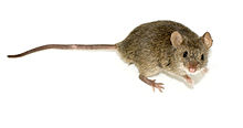
| House mouse (Mus musculus) | |
| Scientific classification | |
| Domain: | Eukaryota |
| Kingdom: | Animalia |
| Phylum: | Chordata |
| Class: | Mammalia |
| Order: | Rodentia |
| Family: | Muridae |
| Tribe: | Murini |
| Genus: | Mus Linnaeus, 1758 |
Introduction
[edit]Mouse development is the process mouse eggs undergo to reach their adult form. This article specifically focuses on embryogenesis. These are some of the earliest stages that initiate development, define early tissue layers, and set body axes. Development during embryogenesis is well conserved. This has led to the use of animals models such as the mouse to study development in humans.
Life Cycle
[edit]Mice are viviparous mammals. Embryogenesis occurs internally following fertilization in females. The fetus continues to develop for an average gestation period of about twenty days.[1] Multiple oocytes may be released during ovulation leading to litter sizes range from about four to twelve pups.[2][3] Each pup is born nude, blind, and earless.[1] Adult mice are sexually mature at 50 days of age with females potentially having their first estrous cycle from twenty-five to forty days of age. Although wild mice may survive to be up to four years of age, the prevalence of predators often shortens the average lifespan to within two years[4].
Fertilization
[edit]
Germ cells undergo meiosis to produce haploid oocytes and sperm cells. Modifications are made within the sperm to prepare for fertilization. These include condensation of the DNA and modifications of organelles. The acrosome develops in front of the nucleus and the flagellum forms behind the nucleus.[5] Egg, or oocyte, cells are arrested in metaphase II of meiosis. Completion of meiosis does not occur until after fertilization [3]. Egg cells are secreted into the oviduct following mating as cumulus cells, oocytes enveloped in a zona pellucida and surrounded by follicle cells [3][6].
Following mating, sperm cells navigate to the oviduct, completing capacitation during this time [3]. Fertilization of the oocyte occurs within the oviduct. Sperm cells bind to the zona pellucida by interacting with ZP3 and ZP2 [3][7]. The initial interaction between the sperm cell and ZP3 activates the acrosomal reaction. The contents of the acrosome are released to digest the zona pellucida [3]. Sperm-egg recognition is carried out at the egg's surface with ADAM and integrin bindings [3]. Research shows there is a preference for binding at the equatorial region, ninety degrees from the location of the first polar body. When the sperm binds, it alters the distribution of transcription factors later in cleavage[8]. Once bound, plasma membranes of both gametes fuse. The contents of the sperm are then integrated into the egg.
After fusion, intracellular calcium levels begin to oscillate and meiosis is resumed. Cortical granules release and migrate to the plasma membrane. These cortical granules then modify receptors to block fusion by more sperm [3]. A second polar body is expelled with the completion of the final meiotic division. DNA replication occurs within the paternal and maternal nucleus. The chromosomes and maternal spindle fibers then align and the zygote is ready for cleavage.[3]
Cleavage
[edit]The developmental step after fertilization is cleavage. The zygote begins division into individual cells. There is no growth involved with these divisions. Forty-eight hours after fertilization, the first three cleavages has occurred. The genes that make up the zygote begin to be expressed once cleavage has resulted in two cells [3]. The individual cells resulting from cleavage are called blastomeres. Once eight cells are reached the process of compaction begins. Compaction is important for increasing intracellular contact. Blastomeres flatten out and the embryo changes shape, looking more spherical. E-cadherin is a molecule that is heavily involved in the process of compaction[9]. Gap junctions are formed during compaction as well, these are necessary for the diffusion of molecules in the embryo [3].
Once compaction is complete the embryo is called a morula and is referred to as so until thirty- two cells have been reached. Cleavage continues, and the embryo will start forming a blastocoel. The blastocoel is a cavity full of fluid that begins forming after thirty-two cells are reached. For formation to begin, desmosomes and tight junctions must be present in the embryo creating a seal between the inside and outside. The embryo moves into the uterus during the process of blastocoel formation [3]. The tight junctions keep the fluid in the cavity as it expands [10]. The blastocoel will continue expanding the embryo, into the blastocyst. The blastocyst is divided into two layers, one inner, the other outer[3]. The outer layer, the trophectoderm, contains tight junctions that help form a protective barrier over the inner layer[11]. The inner layer is made up of the cavity fluid and a ball of cells called the inner cell mass. These newly formed layers will give rise to new tissues. Tissue formed in the inner cell mass is called the primitive endoderm. The trophectoderm forms two tissues, one of which is polar. The other tissue becomes polytene cells. These cells contain DNA which replicates and increase. There is no mitosis involved with this DNA replication [3].
Gastrulation
[edit]The developmental process of gastrulation results in the embryo consisting of three layers, the ectoderm, mesoderm, and endoderm. The embryo at the beginning of gastrulation has a cup like shape. The process of gastrulation begins in the epiblast, specifically the posterior end. This end of the epiblast will give rise to the primitive streak and node. This streak lays out the future anterior-posterior axis of the embryo. The spot where the primitive streak begins forming, marks the posterior end[12]. The streak is a series of cell movements that spreads across the embryo. During these cell movements rapid cell division is occurring[2] .The process of cell movement through the primitive streak is regulated by genes being expressed and cell signaling proteins[12] . A gene called Nodal was identified to be heavily involved with the regulation of the primitive streak as it forms and moves across the embryo[13]The formation of the primitive streak also establishes the future left-right axis[14].
Once the streak spreads across the embryo, the node will be visible in the anterior end. Unlike the streak, which is made up of three layers, the node only has two. The node will move back across the embryo to the posterior end[3]. As it moves it releases two important signaling proteins known as Noggin and Chordin. The cells from the node are important for forming the structures that make up the body of the embryo. One of these structures is the notochord which is involved in process of neurulation[15]. The developmental step following gastrulation is neurulation, which is described below.
Neurulation
[edit]Neurulation is defined as morphogenic movements within the primitive streak to form an enclosed neural tube.[15] Morphogens can be defined as intracellular signals. These signals have the capability to inhibit or activate cell growth. In mice, the primitive streak extension begins on day 6.5.[3] The primitive streak can be defined as cell growth to form the midline anterior-posterior axis of an embryo.[3] Here, anterior and posterior are words that represent the future head and tail sites.
The anterior tip of the embryo denotes where head development will occur. To begin neurulation, the neural plate needs to develop. The neural plate will become the neural tube later on in development. To create the neural tube, the anterior cells within the primitive streak need to replicate. In order for the correct cells to replicate, an important mass of morphogens need to be inhibited.[15] The inhibition morphogens are exported by a signaling center called the node. This correct cell replication causes a thickening in that region. The thickening grows into a U shape. In mice, this occurs on day 7.5 of development.[3]
Next, the U-shaped mass of cells needs to extend. The U-shaped mass becomes thinner and longer within the anterior region. In order to form a closed tube, the U-shaped mass needs to fold. The neural plate U is induced to fold. Morphogens are secreted by the notochord, which lay underneath the neural plate. Hinge regions are created within the neural plate and assist in the fold. The folding occurs during day 9-10 of embryo development.[16] The folding that occurs due to the hinge regions allow both ends of the neural-U, or neural folds, to touch along the dorsal midline of the neural plate.[3] Fusion begins once the neural folds touch. And the neural plate is now a neural tube. The neural tube becomes hollow at the dorsal end of the embryo.[17]
Axis Signaling
[edit]Like coordinates on a map, a developing embryo needs axis. Certain morphogens, or cell signals, direct cells when and where to replicate. Replication of specific cells create a head-tail axis, also known as the anterior-posterior axis; as well as the ventral-dorsal axis and left-right axis. Because embryos grow outwards as well as 2-dimnesionally – 3 axis need to be established. This gradients are typically established by cellular gradients created by morphogens. [18]
Many of the morphens, or signaling cells, are excreted by the node in mice. To help establish the anterior-posterior axis. A posterior (or tail-end) extension signal is excreted, called FGF.[19]
Advantages/Disadvantages
[edit]An advantage to using mice as a model organism for development is that they are easy to breed. They also don’t require a lot of space to maintain housing for them and their pups. Since mice are mammals, studying their development can provide insight to human development [20]. A disadvantage is that fertilization is internal [3]. Another disadvantage is that the embryo develops inside of another organism instead of outside like other animals, who develop externally in their environment [20].
- ^ a b "Mouse". Wikipedia. 2018-03-09.
- ^ "Mouse breeding colony management" (PDF). Purdue University.
- ^ a b c d e f g h i j k l m n o p q r s Slack, JMW, ed. (2013). Essential Developmental Biology (3. Ed.). Wiley-Blackwell: John Wiley & Sons. IBSN 978-0-470-92351-1
- ^ Miller, R. A., Harper, J. M., Dysko, R. C., Durkee, S. J., & Austad, S. N. (2002). Longer life spans and delayed maturation in wild-derived mice. Experimental Biology And Medicine (Maywood, N.J.), 227(7), 500-508.
- ^ Li, Ren-Ke; Tan, Jue-Ling; Chen, Li-Ting; Feng, Jing-Sheng; Liang, Wen-Xue; Guo, Xue-Jiang; Liu, Ping; Chen, Zhu; Sha, Jia-Hao (2014-05-21). "Iqcg Is Essential for Sperm Flagellum Formation in Mice". PLOS ONE. 9 (5): e98053. doi:10.1371/journal.pone.0098053. ISSN 1932-6203. PMC 4029791. PMID 24849454.
- ^ Liu, Yang; Wei, Zhuying; Huang, Yafei; Bai, Chunling; Zan, Linsen; Li, Guangpeng (2014). Cyclopamine did not affect mouse oocyte maturation in vitro but decreased early embryonic development. Animal Science Journal, 85: 840-847.
- ^ Howes, Elizabeth; Pascall, John C.; Engel, Wolfgang; Jones, Roy (2001). Interactions between mouse ZP2 glycoprotein and proacrosin; a mechanism for secondary binding of sperm to the zona pellucida during fertilization. Journal of Cell Science 114: 4127-4136.
- ^ Piotrowska-Nitsche, K.; Chan, A. S. (2013). Effect of sperm entry on blastocyst development after in vitro fertilization and intracytoplasmic sperm injection - mouse model. Journal of Assisted Reproduction and Genetics, 30(1), 81-89. doi:10.1007/s10815-012-9896-6
- ^ Larue, L; Ohsugi, M; Hirchenhain, J; Kemler, R (1994). "E-cadherin null mutant embryos fail to form a trophectoderm epithelium". Proceeding of the National Academy of Sciences of the United States of America. 91 (17): 8263–8267. doi:10.1073/pnas.91.17.8263. PMC 44586. PMID 8058792 – via JSTOR.
- ^ Eckert, J J; McCallum, A; Mears, A; Rumbsy, M G; Cameron, I T; Fleming, T P (2004). "Specific PKC isoforms regulate blastocoel formation during mouse preimplantation development". Developmental Biology. 274 (2): 384–401. doi:10.1016/j.ydbio.2004.07.027. PMID 15385166.
- ^ Moriwaki, K; Tsukita, S; Furuse, M (2007). "Tight junctions containing claudin 4 and 6 are essential for blastocyst formation in preimplantation mouse embryos". Developmental Biology. 312 (2): 509–522. doi:10.1016/j.ydbio.2007.09.049. PMID 17980358. S2CID 21575148.
- ^ a b Slack, JMW, ed. (2013). Essential Developmental Biology (3. Ed.). Wiley-Blackwell: John Wiley & Sons. IBSN 978-0-470-92351-1
- ^ Conlon, F. L.; Lyons, K. M.; Takaesu, N.; Barth, K. S.; Kispert, A.; Herrmann, B.; Robertson, E.J. (1994). "A primary requirement for nodal in the formation and maintenance of the primitive streak in the mouse". Development. 120 (7): 1919–1928. doi:10.1242/dev.120.7.1919. PMID 7924997.
- ^ Tam, P P L (1997). "Mouse gastrulation: the formation of a mammalian body plan". Mechanisms of Development. 68 (1–2): 3–25. doi:10.1016/S0925-4773(97)00123-8. PMID 9431800. S2CID 14052942.
- ^ a b Colas, JF; Schoenwolf, GC; (2001) “Towards a cellular and molecular understanding of neurulation”. Developmental Dynamics, 221: 117-145.
- ^ Ybot-Gonzalez, P; Gaston-Massuet, C; Girdler, G; Klingensmith, J; Arkell, R; Greene, NDE; Copp, AJ; (2007) “Neural plate morphogenesis during mouse neurulation is regulated by antagonism of Bmp signaling”. Development and Disease, 134: 3203-3211. Doi: 10.1242/dev.008177.
- ^ Colas, JF; Schoenwolf, GC; (2001) “Towards a cellular and molecular understanding of neurulation” Developmental Dynamics 145: 117-145
- ^ Naganathan, SR; Oates, AC; (2017) “The sweetness of embryonic elongation and differentiation” Developmental Cell 40: Doi:10.1016/j.devcel.2017.02.012.
- ^ Oginuma, M; Moncuquet, P; Xiong, F; Karoly, E; Chal, J; Guevorkian, K; Pourquie, O; (2017) “A gradient of glycolytic activity coordinates Fgf and Wnt signaling during elongation of the body axis in amniote embryos” Developmental Cell 40: 342-353. Doi:10.1016/j.devcel.2017.02.001.
- ^ a b Gibson, S F (2000). Developmental Biology. Sunderland, MA: Sinauer Associates.
