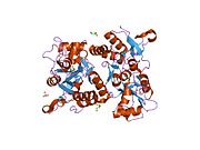
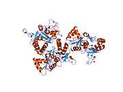
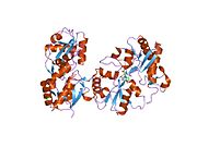
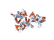
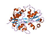
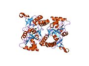
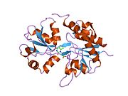
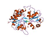
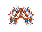
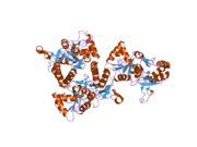
Template:PDB Gallery/2891
Appearance
-
1ftj: CRYSTAL STRUCTURE OF THE GLUR2 LIGAND BINDING CORE (S1S2J) IN COMPLEX WITH GLUTAMATE AT 1.9 RESOLUTION
-
1ftk: CRYSTAL STRUCTURE OF THE GLUR2 LIGAND BINDING CORE (S1S2I) IN COMPLEX WITH KAINATE AT 1.6 A RESOLUTION
-
1ftl: CRYSTAL STRUCTURE OF THE GLUR2 LIGAND BINDING CORE (S1S2J) IN COMPLEX WITH THE ANTAGONIST DNQX AT 1.8 A RESOLUTION
-
1ftm: CRYSTAL STRUCTURE OF THE GLUR2 LIGAND BINDING CORE (S1S2J) IN COMPLEX WITH AMPA AT 1.7 RESOLUTION
-
1fto: CRYSTAL STRUCTURE OF THE GLUR2 LIGAND BINDING CORE (S1S2J) IN THE APO STATE AT 2.0 A RESOLUTION
-
1fw0: CRYSTAL STRUCTURE OF THE GLUR2 LIGAND BINDING CORE (S1S2J) IN COMPLEX WITH KAINATE AT 2.0 A RESOLUTION
-
1gr2: STRUCTURE OF A GLUTAMATE RECEPTOR LIGAND BINDING CORE (GLUR2) COMPLEXED WITH KAINATE
-
1lb8: Crystal structure of the Non-desensitizing GluR2 ligand binding core mutant (S1S2J-L483Y) in complex with AMPA at 2.3 resolution
-
1lb9: Crystal structure of the Non-desensitizing GluR2 ligand binding core mutant (S1S2J-L483Y) in complex with antagonist DNQX at 2.3 A resolution
-
1lbb: Crystal structure of the GluR2 ligand binding domain mutant (S1S2J-N754D) in complex with kainate at 2.1 A resolution
-
1lbc: Crystal structure of GluR2 ligand binding core (S1S2J-N775S) in complex with cyclothiazide (CTZ) as well as glutamate at 1.8 A resolution
-
1m5b: X-RAY STRUCTURE OF THE GLUR2 LIGAND BINDING CORE (S1S2J) IN COMPLEX WITH 2-Me-Tet-AMPA AT 1.85 A RESOLUTION.
-
1m5c: X-RAY STRUCTURE OF THE GLUR2 LIGAND BINDING CORE (S1S2J) IN COMPLEX WITH Br-HIBO AT 1.65 A RESOLUTION
-
1m5d: X-RAY STRUCTURE OF THE GLUR2 LIGAND BINDING CORE (S1S2J-Y702F) IN COMPLEX WITH Br-HIBO AT 1.73 A RESOLUTION
-
1m5e: X-RAY STRUCTURE OF THE GLUR2 LIGAND BINDING CORE (S1S2J) IN COMPLEX WITH ACPA AT 1.46 A RESOLUTION
-
1m5f: X-RAY STRUCTURE OF THE GLUR2 LIGAND BINDING CORE (S1S2J-Y702F) IN COMPLEX WITH ACPA AT 1.95 A RESOLUTION
-
1mm6: crystal structure of the GluR2 ligand binding core (S1S2J) in complex with quisqualate in a non zinc crystal form at 2.15 angstroms resolution
-
1mm7: Crystal Structure of the GluR2 Ligand Binding Core (S1S2J) in Complex with Quisqualate in a Zinc Crystal Form at 1.65 Angstroms Resolution
-
1mqd: X-ray structure of the GluR2 ligand-binding core (S1S2J) in complex with (S)-Des-Me-AMPA at 1.46 A resolution. Crystallization in the presence of lithium sulfate.
-
1mqg: Crystal Structure of the GluR2 Ligand Binding Core (S1S2J) in Complex with Iodo-Willardiine at 2.15 Angstroms Resolution
-
1mqh: Crystal Structure of the GluR2 Ligand Binding Core (S1S2J) in Complex with Bromo-Willardiine at 1.8 Angstroms Resolution
-
1mqi: Crystal Structure of the GluR2 Ligand Binding Core (S1S2J) in Complex with Fluoro-Willardiine at 1.35 Angstroms Resolution
-
1mqj: Crystal structure of the GluR2 ligand binding core (S1S2J) in complex with willardiine at 1.65 angstroms resolution
-
1ms7: X-ray structure of the GluR2 ligand-binding core (S1S2J) in complex with (S)-Des-Me-AMPA at 1.97 A resolution, Crystallization in the presence of zinc acetate
-
1mxu: CRYSTAL STRUCTURE OF THE GLUR2 LIGAND BINDING CORE (S1S2J) in complex with bromo-willardiine (Control for the crystal titration experiments)
-
1mxv: crystal titration experiments (AMPA co-crystals soaked in 10 mM BrW)
-
1mxw: crystal titration experiments (AMPA co-crystals soaked in 1 mM BrW)
-
1mxx: crystal titration experiments (AMPA co-crystals soaked in 100 uM BrW)
-
1mxy: crystal titration experiments (AMPA co-crystals soaked in 10 uM BrW)
-
1mxz: crystal titration experiments (AMPA co-crystals soaked in 1 uM BrW)
-
1my0: crystal titration experiments (AMPA co-crystals soaked in 100 nM BrW)
-
1my1: crystal titration experiments (AMPA co-crystals soaked in 10 nM BrW)
-
1my2: crystal titration experiment (AMPA complex control)
-
1my3: crystal structure of glutamate receptor ligand-binding core in complex with bromo-willardiine in the Zn crystal form
-
1my4: crystal structure of glutamate receptor ligand-binding core in complex with iodo-willardiine in the Zn crystal form
-
1n0t: X-ray structure of the GluR2 ligand-binding core (S1S2J) in complex with the antagonist (S)-ATPO at 2.1 A resolution.
-
1nnk: X-ray structure of the GluR2 ligand-binding core (S1S2J) in complex with (S)-ATPA at 1.85 A resolution. Crystallization with zinc ions.
-
1nnp: X-ray structure of the GluR2 ligand-binding core (S1S2J) in complex with (S)-ATPA at 1.9 A resolution. Crystallization without zinc ions.
-
1p1n: GluR2 Ligand Binding Core (S1S2J) Mutant L650T in Complex with Kainate
-
1p1o: Crystal structure of the GluR2 ligand-binding core (S1S2J) mutant L650T in complex with quisqualate
-
1p1q: Crystal structure of the GluR2 ligand binding core (S1S2J) L650T mutant in complex with AMPA
-
1p1u: Crystal structure of the GluR2 ligand-binding core (S1S2J) L650T mutant in complex with AMPA (ammonium sulfate crystal form)
-
1p1w: Crystal structure of the GluR2 ligand-binding core (S1S2J) with the L483Y and L650T mutations and in complex with AMPA
-
1syh: X-RAY STRUCTURE OF THE GLUR2 LIGAND-BINDING CORE (S1S2J) IN COMPLEX WITH (S)-CPW399 AT 1.85 A RESOLUTION.
-
1syi: X-RAY STRUCTURE OF THE Y702F MUTANT OF THE GLUR2 LIGAND-BINDING CORE (S1S2J) IN COMPLEX WITH (S)-CPW399 AT 2.1 A RESOLUTION.
-
1wvj: Exploring the GluR2 ligand-binding core in complex with the bicyclic AMPA analogue (S)-4-AHCP
-
1xhy: X-ray structure of the Y702F mutant of the GluR2 ligand-binding core (S1S2J) in complex with kainate at 1.85 A resolution
-
2aix: X-ray structure of the GLUR2 ligand-binding core (S1S2J) in complex with (s)-thio-atpa at 2.2 a resolution.
-
2al4: CRYSTAL STRUCTURE OF THE GLUR2 LIGAND BINDING CORE (S1S2J) IN COMPLEX WITH quisqualate and CX614.
-
2al5: Crystal structure of the GluR2 ligand binding core (S1S2J) in complex with fluoro-willardiine and aniracetam
-
2anj: Crystal Structure of the Glur2 Ligand Binding Core (S1S2J-Y450W) Mutant in Complex With the Partial Agonist Kainic Acid at 2.1 A Resolution
-
2cmo: THE STRUCTURE OF A MIXED GLUR2 LIGAND-BINDING CORE DIMER IN COMPLEX WITH (S)-GLUTAMATE AND THE ANTAGONIST (S)-NS1209
-
2gfe: Crystal structure of the GluR2 A476E S673D Ligand Binding Core Mutant at 1.54 Angstroms Resolution
-
2i3v: Measurement of conformational changes accompanying desensitization in an ionotropic glutamate receptor: Structure of G725C mutant
-
2i3w: Measurement of conformational changes accompanying desensitization in an ionotropic glutamate receptor: Structure of S729C mutant
-
2uxa: CRYSTAL STRUCTURE OF THE GLUR2-FLIP LIGAND BINDING DOMAIN, R/G UNEDITED.


