Genital tubercle
| Genital tubercle | |
|---|---|
 Stages in the development of the external sexual organs in the male and female. | |
| Details | |
| Precursor | Somatopleure[1] |
| Gives rise to | Clitoris or penis |
| System | Reproductive system |
| Identifiers | |
| Latin | tuberculum phallicum; tuberculum genitale |
| TE | tubercle_by_E5.7.4.0.1.0.1 E5.7.4.0.1.0.1 |
| Anatomical terminology | |
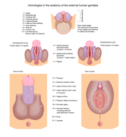
A genital tubercle, phallic tubercle, or clitorophallic structure[2] is a body of tissue present in the development of the reproductive system of amniotes.[3] It forms in the ventral, caudal region of mammalian embryos of both sexes, and eventually develops into a primordial phallus. In the human fetus, the genital tubercle develops around week four of gestation, and by week nine, becomes recognizably either a clitoris or penis. This should not be confused with the sinus tubercle which is a proliferation of endoderm induced by paramesonephric ducts. Even after the phallus is developed (either a penile shaft or clitoral shaft),[4] the term genital tubercle remains, but only as the terminal end of it,[5] which develops into either the glans penis or the glans clitoridis.
In the development of the male fetus, the two sides of the tubercle approach ventrally forming a hollow tube that encloses the male urethra. The two glans wings merge in the midline forming the septum glandis.[6] In the female fetus, the tubercle is attached to the vestibular folds that remain unfused forming the labia minora and the vaginal vestibule in between.[7] The genital tubercle is sensitive to dihydrotestosterone and rich in 5-alpha-reductase, so that the amount of fetal testosterone present after the second month is a major determinant of phallus size at birth.
See also
[edit]References
[edit]- ^ Netter, Frank H.; Cochard, Larry R. (2002). Netter's Atlas of human embryology. Teterboro, N.J: Icon Learning Systems. p. 159. ISBN 0-914168-99-1.
- ^ Skinner, Michael K. (2018). Encyclopedia of Reproduction: Volume 1. Elsevier Science. p. 445. ISBN 978-0-12815-145-7.
- ^ Prum, Richard O. (2023). Performance All the Way Down: Genes, Development, and Sexual Difference. University of Chicago Press. p. 219. ISBN 978-0-22682-978-4.
- ^ Netter, Frank (2022). Netter Atlas of Human Anatomy: Classic Regional Approach - Ebook. Elsevier Health Sciences. p. 194. ISBN 978-0-32379-375-9. Retrieved October 9, 2023.
- ^ "Urogenital Development - External Genitalia 24 of 28". Units of Embryo Images. UNC School of Medicine. Archived from the original on Apr 11, 2011.
- ^ Joseph, Diya B.; Vezina, Chad M. (2018), "Male Reproductive Tract: Development Overview", Encyclopedia of Reproduction, Elsevier, pp. 248–255, doi:10.1016/B978-0-12-801238-3.64366-0, ISBN 9780128151457, retrieved 2023-01-04
- ^ Baskin, Laurence; Shen, Joel; Sinclair, Adriane; Cao, Mei; Liu, Xin; Liu, Ge; Isaacson, Dylan; Overland, Maya; Li, Yi; Cunha, Gerald R. (2018). "Development of the human penis and clitoris". Differentiation; Research in Biological Diversity. 103: 74–85. doi:10.1016/j.diff.2018.08.001. ISSN 1432-0436. PMC 6234061. PMID 30249413.
External links
[edit]- Swiss embryology (from UL, UB, and UF) ugenital/genitexterne01
- Overview at mcgill.ca
