Ribokinase
| ribokinase | |||||||||
|---|---|---|---|---|---|---|---|---|---|
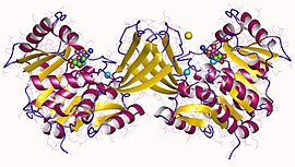
 Ribokinase dimer, Human | |||||||||
| Identifiers | |||||||||
| EC no. | 2.7.1.15 | ||||||||
| CAS no. | 9026-84-0 | ||||||||
| Databases | |||||||||
| IntEnz | IntEnz view | ||||||||
| BRENDA | BRENDA entry | ||||||||
| ExPASy | NiceZyme view | ||||||||
| KEGG | KEGG entry | ||||||||
| MetaCyc | metabolic pathway | ||||||||
| PRIAM | profile | ||||||||
| PDB structures | RCSB PDB PDBe PDBsum | ||||||||
| Gene Ontology | AmiGO / QuickGO | ||||||||
| |||||||||
In enzymology, a ribokinase (EC 2.7.1.15) is an enzyme that catalyzes the chemical reaction
- ATP + d-ribose ⇌ ADP + d-ribose 5-phosphate
Thus, the two substrates of this enzyme are ATP and d-ribose, whereas its two products are ADP and d-ribose 5-phosphate.
The systematic name of this enzyme class is ATP:d-ribose 5-phosphotransferase. Other names in common use include deoxyribokinase, ribokinase (phosphorylating), and d-ribokinase. This enzyme participates in pentose phosphate pathway.
Ribokinase (RK) belongs to the phosphofructokinase B (PfkB) family of sugar kinases.[1] Other members of this family (also known as the RK family) include adenosine kinase (AK), inosine-guanosine kinase, fructokinase, and 1-phosphofructokinase.[1][2][3] The members of the PfkB/RK family are identified by the presence of three conserved sequence motifs and the enzymatic activity of this family of protein generally shows a dependence on the presence of pentavalent ions.[1][2][4] The conserved NXXE motif, which is a distinctive property of the PfkB family of proteins, is involved in pentavalent ion dependency. The structures of RK and several other PfK family of proteins have been determined from a number of organisms.[5] Despite low sequence similarity between AdK and other PfkB family of proteins, these proteins are quite similar at structural levels.[1]
Structural studies
[edit]As of late 2007, 7 structures have been solved for this class of enzymes, with PDB accession codes 1GQT, 1RK2, 1RKA, 1RKD, 1RKS, 1VM7, and 2FV7.
References
[edit]- ^ a b c d Park J, Gupta RS (September 2008). "Adenosine kinase and ribokinase--the RK family of proteins". Cellular and Molecular Life Sciences. 65 (18): 2875–96. doi:10.1007/s00018-008-8123-1. PMC 11131688. PMID 18560757. S2CID 11439854.
- ^ a b Bork P, Sander C, Valencia A (January 1993). "Convergent evolution of similar enzymatic function on different protein folds: the hexokinase, ribokinase, and galactokinase families of sugar kinases". Protein Science. 2 (1): 31–40. doi:10.1002/pro.5560020104. PMC 2142297. PMID 8382990.
- ^ Spychala J, Datta NS, Takabayashi K, Datta M, Fox IH, Gribbin T, Mitchell BS (February 1996). "Cloning of human adenosine kinase cDNA: sequence similarity to microbial ribokinases and fructokinases". Proceedings of the National Academy of Sciences of the United States of America. 93 (3): 1232–7. Bibcode:1996PNAS...93.1232S. doi:10.1073/pnas.93.3.1232. PMC 40062. PMID 8577746.
- ^ Maj MC, Singh B, Gupta RS (March 2002). "Pentavalent ions dependency is a conserved property of adenosine kinase from diverse sources: identification of a novel motif implicated in phosphate and magnesium ion binding and substrate inhibition". Biochemistry. 41 (12): 4059–69. doi:10.1021/bi0119161. PMID 11900549.
- ^ Sigrell JA, Cameron AD, Jones TA, Mowbray SL (February 1998). "Structure of Escherichia coli ribokinase in complex with ribose and dinucleotide determined to 1.8 A resolution: insights into a new family of kinase structures". Structure. 6 (2): 183–93. doi:10.1016/s0969-2126(98)00020-3. PMID 9519409.
Further reading
[edit]- Agranoff BW, Brady RO (March 1956). "Purification and properties of calf liver ribokinase" (PDF). The Journal of Biological Chemistry. 219 (1): 221–9. doi:10.1016/S0021-9258(18)65786-2. PMID 13295274.
- Ginsburg A (March 1959). "A deoxyribokinase from Lactobacillus plantarum" (PDF). The Journal of Biological Chemistry. 234 (3): 481–7. doi:10.1016/S0021-9258(18)70230-5. PMID 13641245.
