Transrectal ultrasonography
Appearance
(Redirected from Transrectal ultrasound)
| Transrectal ultrasonography | |
|---|---|
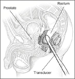
 A probe inserted in the rectum emits sound waves to image the prostate | |
| ICD-9-CM | 88.74 |
| OPS-301 code | 3-058 |
Transrectal ultrasonography, or TRUS in short, is a method of creating an image of organs in the pelvis, most commonly used to perform an ultrasound-guided needle biopsy evaluation of the prostate gland in men with elevated prostate-specific antigen or prostatic nodules on digital rectal exam. TRUS-guided biopsy may reveal prostate cancer, benign prostatic hyperplasia, or prostatitis. TRUS may also detect other diseases of the lower rectum and can be used to stage primary rectal cancer.[1][2][3]
References
[edit]- ^ O' Donoghue PM, McSweeney SE, Jhaveri K (2010). "Genitourinary imaging: current and emerging applications". J Postgrad Med. 56 (2): 131–9. doi:10.4103/0022-3859.65291. PMID 20622393.
- ^ Shetty, Sugandh (4 August 2016). "Transrectal Ultrasonography of the Prostate". Medscape. Retrieved 18 June 2019.(subscription required)
- ^ Kim, Min Ju (2014-11-19). "Transrectal ultrasonography of anorectal diseases: advantages and disadvantages". Ultrasonography. 34 (1): 19–31. doi:10.14366/usg.14051. ISSN 2288-5919. PMC 4282231. PMID 25492891.
