Sinoatrial nodal artery
| Sinoatrial nodal artery | |
|---|---|
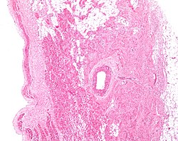
 Low magnification micrograph of a sinoatrial node (central on image), surrounding the sinuatrial nodal artery (on lumen in the image). H&E stain. | |
 Arteries: RCA = right coronary AB = atrial branches SANB = sinuatrial nodal RMA = right marginal LCA = left coronary CB = circumflex branch LAD/AIB = anterior interventricular LMA = left marginal PIA/PDA = posterior descending MARG = left marginal AVN = atrioventricular nodal Veins: SCV = small cardiac ACV = anterior cardiac AIV/GCV = great cardiac MCV = middle cardiac CS = coronary sinus | |
| Details | |
| Identifiers | |
| Latin | ramus nodi sinuatrialis arteriae coronariae dextrae |
| TA98 | A12.2.03.104 |
| TA2 | 4133 |
| FMA | 3823 |
| Anatomical terminology | |

The sinoatrial nodal artery, sinoatrial nodal artery or sinoatrial artery is an artery of the heart which supplies the sinoatrial node, the natural pacemaker center of the heart. It is usually a branch of the right coronary artery.[1] It passes between the right atrium, and the opening of the superior vena cava.[2]
Anatomy
[edit]Origin
[edit]It arises from the right coronary artery in around 60% of individuals, from the left circumflex coronary artery in about 40% of individuals,[1] and in less than 1% of humans, the artery has an anomalous origin directly from the coronary sinus, descending aorta, or distal right coronary artery.[citation needed]
The origin of the sinoatrial node artery is not related to coronary artery dominance, which means the side (right or left) that provides the circulation to the back of the heart. In contrast, the atrioventricular nodal branch, that is the artery that brings blood to the atrioventricular node, depends on coronary artery dominance.[citation needed]
Course
[edit]In more than 50% of human hearts, the artery actually courses close to the superior posterior aspect of the interatrial septum.[3]
The sinoatrial node surrounds the sinoatrial artery, which can run centrally (in 70% of individuals) or off-center within the node.[4]
Variation
[edit]A left S-shaped sinoatrial artery, originating from the proximal left circumflex or LCx artery, has been described as a common variant in approximately 10% of human hearts.[5] This artery is larger than normal and supplies a good part of the left atrium, but also right-sided structures like part of the sinoatrial node and the atrioventricular nodal areas. In this variant, the artery courses in the sulcus between the left superior pulmonary vein and the left atrial appendage where it could be susceptible to injury during catheter or surgical ablation procedures on the left atrium, especially for atrial fibrillation ablation or open-heart cardiac surgery.[citation needed]
Additional images
[edit]
-
Sternocostal surface of heart (right coronary artery visible at left)
References
[edit]- ^ a b Pejković, B; Krajnc, I; et al. (July 2008). "Anatomical Aspects of the Arterial Blood Supply to the Sinoatrial and Atrioventricular Nodes of the Human Heart". Journal of International Medical Research. 36 (4): 691–698. doi:10.1177/147323000803600410. PMID 18652764.
- ^ Morton, David A. (2018). The Big Picture: Gross Anatomy. K. Bo Foreman, Kurt H. Albertine (2nd ed.). New York. p. 54. ISBN 978-1-259-86264-9. OCLC 1044772257.
{{cite book}}: CS1 maint: location missing publisher (link) - ^ Click, Roger L.; Holmes, David R.; et al. (March 1989). "Anomalous coronary arteries: location, degree of atherosclerosis and effect on survival—a report from the coronary artery surgery study". Journal of the American College of Cardiology. 13 (3): 531–537. doi:10.1016/0735-1097(89)90588-3. PMID 2918156.
- ^ Sánchez-Quintana, D.; Anderson, R. H.; et al. (1 October 2002). "The terminal crest: morphological features relevant to electrophysiology". Heart. 88 (4): 406–411. doi:10.1136/heart.88.4.406. PMC 1767383. PMID 12231604.
- ^ Saremi, Farhood; Channual, Stephanie; et al. (June 2008). "MDCT of the S-Shaped Sinoatrial Node Artery". American Journal of Roentgenology. 190 (6): 1569–1575. doi:10.2214/AJR.07.3127. PMID 18492908.

