
Pineal gland: Difference between revisions
Undid revision 280786310 by 76.126.82.26 (talk) |
No edit summary |
||
| Line 22: | Line 22: | ||
==Location== |
==Location== |
||
The pineal gland is reddish-gray and about the size of a [[pea]] (8 mm in humans), located just rostro-dorsal to the [[superior colliculus]] and behind and beneath the [[stria medullaris]], between the laterally positioned [[thalamic bodies]]. It is part of the [[epithalamus]]. |
The pineal gland is reddish-gray and about the size of a [[pea]] (8 mm in humans), located just rostro-dorsal to the [[superior colliculus]] and behind and beneath the [[stria medullaris]], between the laterally positioned [[thalamic bodies]]. It is part of the [[epithalamus]].near the middle |
||
The pineal gland is a midline structure, and is often seen in plain [[skull]] [[X-ray]]s, as it is often [[calcification|calcified]]. Calcification is typically due to intake of the [[fluoride]] found in water and toothpaste.{{Fact|date=March 2009}} |
The pineal gland is a midline structure, and is often seen in plain [[skull]] [[X-ray]]s, as it is often [[calcification|calcified]]. Calcification is typically due to intake of the [[fluoride]] found in water and toothpaste.{{Fact|date=March 2009}} |
||
Revision as of 16:16, 31 March 2009
| Pineal gland | |
|---|---|
 Endocrine system | |
 Diagram of pituitary and pineal glands. | |
| Details | |
| Artery | superior cerebellar artery |
| Identifiers | |
| Latin | glandula pinealis |
| MeSH | D010870 |
| NeuroNames | 297 |
| NeuroLex ID | birnlex_1184 |
| TA98 | A11.2.00.001 |
| TA2 | 3862 |
| FMA | 62033 |
| Anatomical terminology | |
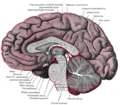
The pineal gland (also called the pineal body, epiphysis cerebri, epiphysis or the "third eye") is a small endocrine gland in the vertebrate brain. It produces melatonin, a hormone that affects the modulation of wake/sleep patterns and photoperiodic (seasonal) functions.[1][2] It is shaped like a tiny pine cone (hence its name), and is located near to the center of the brain, between the two hemispheres, tucked in a groove where the two rounded thalamic bodies join. Unlike much of the rest of the brain, the pineal gland is not isolated from the body by the blood-brain barrier system.[3]
Location
The pineal gland is reddish-gray and about the size of a pea (8 mm in humans), located just rostro-dorsal to the superior colliculus and behind and beneath the stria medullaris, between the laterally positioned thalamic bodies. It is part of the epithalamus.near the middle
The pineal gland is a midline structure, and is often seen in plain skull X-rays, as it is often calcified. Calcification is typically due to intake of the fluoride found in water and toothpaste.[citation needed]
Structure and composition
The pineal body consists in humans of a lobular parenchyma of pinealocytes surrounded by connective tissue spaces. The gland's surface is covered by a pial capsule.
The pineal gland consists mainly of pinealocytes, but four other cell types have been identified.
| Cell type | Description |
| pinealocytes | The pinealocytes consist of a cell body with 4-6 processes emerging. They produce and secrete melatonin. The pinealocytes can be stained by special silver impregnation methods. |
| interstitial cells | Interstitial cells are located between the pinealocytes. |
| perivascular phagocyte | Many capillaries are present in the gland, and perivascular phagocytes are located close to these blood vessels. The perivascular phagocytes are antigen presenting cells. |
| pineal neurons | In higher vertebrates neurons are located in the pineal gland. However, these are not present in rodents. |
| peptidergic neuron-like cells | In some species, neuronal-like peptidergic cells are present. These cells might have a paracrine regulatory function. |
The pineal gland receives a sympathetic innervation from the superior cervical ganglion. However, a parasympathetic innervation from the sphenopalatine and otic ganglia is also present. Further, some nerve fibers penetrate into the pineal gland via the pineal stalk (central innervation). Finally, neurons in the trigeminal ganglion innervates the gland with nerve fibers containing the neuropeptide, PACAP. Human follicles contain a variable quantity of gritty material, called corpora arenacea (or "acervuli", or "brain sand"). Chemical analysis shows that they are composed of calcium phosphate, calcium carbonate, magnesium phosphate, and ammonium phosphate.[4] Recently, calcite deposits have been described as well.[5] Calcium and phosphorus deposits in the pineal gland have been linked with aging.[6]
Miscellaneous anatomy
Pinealocytes in many non-mammalian vertebrates have a strong resemblance to the photoreceptor cells of the eye. Some evolutionary biologists believe that the vertebrate pineal cells share a common evolutionary ancestor with retinal cells.[7]
In some vertebrates, exposure of the pineal to light can directly set off a chain reaction of enzymatic events which regulate circadian rhythms.[8] Some early vertebrate fossil skulls have a pineal foramen (opening). This corroborates with the physiology of the modern "living fossils", the lamprey and the tuatara, and some other vertebrates which have a parietal organ or "third eye" which, in some of them, is photosensitive. The third eye represents evolution’s earlier approach to photoreception.[9] The structures of the third eye in the tuatara are homologous to the cornea, lens and retina, though the latter resembles that of an octopus rather than a vertebrate retina. The asymmetrical whole consists of the "eye" to the left and the pineal sac to the right. "In animals that have lost the parietal eye, including mammals, the pineal sac is retained and condensed into the form of the pineal gland."[9]
Fossils seldom preserve soft anatomy. The brain of the Russian Melovatka bird, about 90 million years old, is an exception, and it shows a larger-than-expected parietal eye and pineal gland.[10]
In humans and other mammals, the light signals necessary to set circadian rhythms are sent from the eye through the retinohypothalamic system to the suprachiasmatic nuclei (SCN) and the pineal.
Function
The pineal gland was originally believed to be a "vestigial remnant" of a larger organ (much as the appendix was thought to be a vestigial digestive organ). Aaron Lerner and colleagues at Yale University discovered that melatonin, the most potent compound then known to lighten frog skin, was present in the highest concentrations in the pineal.[11] Melatonin is a derivative of the amino acid tryptophan, which also has other functions in the central nervous system. The production of melatonin by the pineal gland is stimulated by darkness and inhibited by light.[12] Photosensitive cells in the retina detect light and directly signal the suprachiasmatic nucleus (SCN), entraining its rhythm to the 24-hour cycle in nature. Fibers project from the SCN to the paraventricular nuclei (PVN), which relay the circadian signals to the spinal cord and out via the sympathetic system to superior cervical ganglia (SCG), and from there into the pineal gland. The function(s) of melatonin in humans is not clear; it is commonly prescribed for the treatment of circadian rhythm sleep disorders.
The compound pinoline is also produced in the pineal gland; it is one of the beta-carbolines.
The human pineal gland grows in size until about 1–2 years of age, remaining stable thereafter[13] [14], although its weight increases gradually from puberty onwards [15][16]. The abundant melatonin levels in children is believed to inhibit sexual development, and pineal tumors have been linked with precocious puberty. When puberty arrives, melatonin production is reduced. Calcification of the pineal gland is typical in adults.
In animals, the pineal gland appears to play a major role in sexual development, hibernation, metabolism, and seasonal breeding.
Pineal cytostructure seems to have evolutionary similarities to the retinal cells of chordates.[17] Modern birds and reptiles have been found to express the phototransducing pigment melanopsin in the pineal gland. Avian pineal glands are believed to act like the suprachiasmatic nucleus in mammals.[18]
Studies suggest that in rodents the pineal gland may influence the actions of recreational drugs, such as cocaine,[19] and antidepressants, such as fluoxetine (Prozac),[20] and its hormone melatonin can protect against neurodegeneration.[21]
Cultures, philosophies and mythologies
The secretory activity of the pineal gland has only relatively recently become understood. Historically, its location deep in the brain suggested to philosophers that it possessed particular importance. This combination led to its being a "mystery" gland with myth, superstition and metaphysical theories surrounding its perceived function.
René Descartes, who dedicated much time to the study of the pineal gland,[22] called it the "seat of the soul".[23] He believed that it was the point of connection between the intellect and the body.[24]
The notion of a 'pineal-eye' is central to the philosophy of the seminal French writer Georges Bataille, which is analyzed at length by literary scholar Denis Hollier in his study Against Architecture.[25] In this work Hollier discusses how Bataille uses the concept of a 'pineal-eye' as a reference to a blind-spot in Western rationality.
Additional images
-
 Mesal aspect of a brain sectioned in the median sagittal plane.
 -
 Dissection showing the ventricles of the brain.
 -
 Hind- and mid-brains; postero-lateral view.
 -
 Median sagittal section of brain.


References
- ^ Macchi M, Bruce J (2004). "Human pineal physiology and functional significance of melatonin". Front Neuroendocrinol. 25 (3–4): 177–95. doi:10.1016/j.yfrne.2004.08.001. PMID 15589268.
- ^ Arendt J, Skene DJ (2005). "Melatonin as a chronobiotic". Sleep Med Rev. 9 (1): 25–39. doi:10.1016/j.smrv.2004.05.002. PMID 15649736.
Exogenous melatonin has acute sleepiness-inducing and temperature-lowering effects during 'biological daytime', and when suitably timed (it is most effective around dusk and dawn) it will shift the phase of the human circadian clock (sleep, endogenous melatonin, core body temperature, cortisol) to earlier (advance phase shift) or later (delay phase shift) times.
- ^ Pritchard, Thomas C. (1999). Medical Neuroscience (Google books preview). Hayes Barton Press. pp. 76–77. ISBN 1889325295. Retrieved 2009-02-08.
{{cite book}}: Cite has empty unknown parameters:|origmonth=,|month=,|chapterurl=, and|origdate=(help); Unknown parameter|coauthors=ignored (|author=suggested) (help) - ^ Bocchi G, Valdre G (1993). "Physical, chemical, and mineralogical characterization of carbonate-hydroxyapatite concretions of the human pineal gland". J Inorg Biochem. 49 (3): 209–20. doi:10.1016/0162-0134(93)80006-U. PMID 8381851.
- ^ Baconnier S, Lang S, Polomska M, Hilczer B, Berkovic G, Meshulam G (2002). "Calcite microcrystals in the pineal gland of the human brain: first physical and chemical studies". Bioelectromagnetics. 23 (7): 488–95. doi:10.1002/bem.10053. PMID 12224052.
{{cite journal}}: CS1 maint: multiple names: authors list (link) - ^ http://www.ingentaconnect.com/content/hum/bter/2007/00000119/00000002/art00004
- ^ Klein D (2004). "The 2004 Aschoff/Pittendrigh lecture: Theory of the origin of the pineal gland--a tale of conflict and resolution". J Biol Rhythms. 19 (4): 264–79. doi:10.1177/0748730404267340. PMID 15245646.
- ^ Moore RY, Heller A, Wurtman RJ, Axelrod J. Visual pathway mediating pineal response to environmental light. Science 1967;155(759):220–3. PMID 6015532
- ^ a b Schwab, I. R. (2005). "The lonely eye" (Full text). British Journal of Ophthalmology. 89 (3): 256. doi:10.1136/bjo.2004.059105. Retrieved 2009-02-14.
{{cite journal}}: Cite has empty unknown parameters:|laydate=,|laysource=, and|laysummary=(help); Unknown parameter|coauthors=ignored (|author=suggested) (help); Unknown parameter|month=ignored (help) - ^ Kurochkin, Evgeny N. (2007). "A fossil brain from the Cretaceous of European Russia and avian sensory evolution" (Full text). Biology Letters. 3 (3). The Royal Society: 309–313. doi:10.1098/rsbl.2006.0617. Retrieved 2009-02-14.
{{cite journal}}: Unknown parameter|coauthors=ignored (|author=suggested) (help); Unknown parameter|month=ignored (help) - ^ Lerner AB, Case JD, Takahashi Y (1960). "Isolation of melatonin and 5-methoxyindole-3-acetic acid from bovine pineal glands". J Biol Chem. 235: 1992–7. PMID 14415935.
{{cite journal}}: CS1 maint: multiple names: authors list (link) - ^ Axelrod J (1970). "The pineal gland". Endeavour. 29 (108): 144–8. PMID 4195878.
- ^ Lack of pineal growth during childhood. Schmidt F, Penka B, Trauner M, Reinsperger L, Ranner G, Ebner F, Waldhauser F. J Clin Endocrinol Metab. 1995 Apr;80(4):1221-5.
- ^ Development of the pineal gland: measurement with MR. Sumida M, Barkovich AJ, Newton TH. AJNR Am J Neuroradiol. 1996 Feb;17(2):233-6.
- ^ Tapp E, Huxley M. The weight and degree of calcification of the pineal gland. J Pathol 1971;105:31–39
- ^ Tapp E, Huxley M. The histological appearance of the human pineal gland from puberty to old age. J Pathol 1972;108:137–144
- ^ Klein D (2004). "The 2004 Aschoff/Pittendrigh lecture: Theory of the origin of the pineal gland--a tale of conflict and resolution". J Biol Rhythms. 19 (4): 264–79. doi:10.1177/0748730404267340. PMID 15245646.
- ^ Natesan A, Geetha L, Zatz M (2002). "Rhythm and soul in the avian pineal". Cell Tissue Res. 309 (1): 35–45. doi:10.1007/s00441-002-0571-6. PMID 12111535.
{{cite journal}}: CS1 maint: multiple names: authors list (link) - ^ Uz T, Akhisaroglu M, Ahmed R, Manev H (2003). "The pineal gland is critical for circadian Period1 expression in the striatum and for circadian cocaine sensitization in mice". Neuropsychopharmacology. 28 (12): 2117–23. doi:10.1038/sj.npp.1300254. PMID 12865893.
{{cite journal}}: CS1 maint: multiple names: authors list (link) - ^ Uz T, Dimitrijevic N, Akhisaroglu M, Imbesi M, Kurtuncu M, Manev H (2004). "The pineal gland and anxiogenic-like action of fluoxetine in mice". Neuroreport. 15 (4): 691–4. doi:10.1097/00001756-200403220-00023. PMID 15094477.
{{cite journal}}: CS1 maint: multiple names: authors list (link) - ^ Manev H, Uz T, Kharlamov A, Joo J (1996). "Increased brain damage after stroke or excitotoxic seizures in melatonin-deficient rats". FASEB J. 10 (13): 1546–51. PMID 8940301.
{{cite journal}}: CS1 maint: multiple names: authors list (link) - ^ Descartes and the Pineal Gland (Stanford Encyclopedia of Philosophy)
- ^ Descartes R. Treatise of Man. New York: Prometheus Books; 2003. ISBN 1-59102-090-5
- ^ Descartes R. "The Passions of the Soul" excerpted from "Philosophy of the Mind", Chalmers, D. New York: Oxford University Press, Inc.; 2002. ISBN-13 978-0-19-514581-6
- ^ Hollier, D, Against Architecture: The Writings of Georges Bataille, trans. Betsy Wing, MIT, 1989.
External links
- Stained brain slice images which include the "pineal%20gland" at the BrainMaps project
- hier-280 at NeuroNames
- Histology image: 14401loa – Histology Learning System at Boston University - "Endocrine System: pineal gland "
- Anatomy Atlases – Microscopic Anatomy, plate 15.296
- Images of Pineal region http://rad.usuhs.edu/medpix/medpix.html?mode=image_finder&action=search&srchstr=pineal&srch_type=all#top
- http://isc.temple.edu/neuroanatomy/lab/atlas/pdhn/