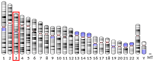
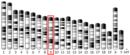
TOPBP1
| TOPBP1 | |||||||||||||||||||||||||||||||||||||||||||||||||||
|---|---|---|---|---|---|---|---|---|---|---|---|---|---|---|---|---|---|---|---|---|---|---|---|---|---|---|---|---|---|---|---|---|---|---|---|---|---|---|---|---|---|---|---|---|---|---|---|---|---|---|---|
 | |||||||||||||||||||||||||||||||||||||||||||||||||||
| Identifiers | |||||||||||||||||||||||||||||||||||||||||||||||||||
| Aliases | TOPBP1, TOP2BP1, topoisomerase (DNA) II binding protein 1, DNA topoisomerase II binding protein 1, Dpb11 | ||||||||||||||||||||||||||||||||||||||||||||||||||
| External IDs | OMIM: 607760; MGI: 1920018; HomoloGene: 38262; GeneCards: TOPBP1; OMA:TOPBP1 - orthologs | ||||||||||||||||||||||||||||||||||||||||||||||||||
| |||||||||||||||||||||||||||||||||||||||||||||||||||
| |||||||||||||||||||||||||||||||||||||||||||||||||||
| |||||||||||||||||||||||||||||||||||||||||||||||||||
| |||||||||||||||||||||||||||||||||||||||||||||||||||
| |||||||||||||||||||||||||||||||||||||||||||||||||||
| Wikidata | |||||||||||||||||||||||||||||||||||||||||||||||||||
| |||||||||||||||||||||||||||||||||||||||||||||||||||
DNA topoisomerase 2-binding protein 1 (TOPBP1) is a scaffold protein that in humans is encoded by the TOPBP1 gene.[5][6][7]
TOPBP1 was first identified as a protein binding partner of DNA topoisomerase-IIβ by a yeast 2-hybrid screen, giving it its name.[8] TOPBP1 is involved in a variety of nuclear specific events. These include DNA damage repair, DNA replication, transcriptional regulation, and cell cycle checkpoint activation. TOPBP1 primarily regulates the DNA damage repair response through its ability to activate the damage response kinase, ataxia-telangiectasia mutated and RAD3-related (ATR). It also plays a critical role in DNA replication initiation and regulation of the cell cycle. Changes in TOPBP1 gene expression are associated with pulmonary hypertension, breast cancer, glioblastoma, non-small cell lung cancer, and sarcomas.[9][10][11]
Structure
[edit]
BRCT domains
[edit]The TOPBP1 gene encodes a scaffold protein which facilitates interactions between different proteins at specific times and locations. It accomplishes these interactions with other protein partners through its breast cancer associated gene 1 C-terminus (BRCT) domains.[10] A BRCT domain is structurally defined by a 4 member β sheet that is bookended by one α-helix (α2) and two other α-helices (α1 and α3). The amino acid residues that make up these core features are highly conserved, with protein specific deviations occurring in the loops that connect these subunits.[13][14] BRCT domains canonically act in pairs, with one domain acting as the acceptor for phosphorylated binding partners and the other domain possessing a binding motif that provides specificity. These pairs are separated by a linker sequence that varies by protein. The paired domains associate through hydrophobic packing interactions that occur between the N-terminal BRCT domain's α2 helix and the C-terminal BRCT domain's α1 and α3 helices. These interactions facilitate BRCT domain binding with phosphorylated binding partners.[13] In contrast, BRCT domains can also exist as either single domains or as a fusion of two different domains.
Human TOPBP1 has nine unique BRCT domains, with four conserved from the budding yeast homologue Dpb11 (i.e. BRCT1,2 and BRCT4,5).[15] In human TOPBP1 the BRCT0, BRCT1, and BRCT2 domains uniquely exist in triple domain form, which is in contrast to the yeast Dpb11 canonical double domain. Only the BRCT3 and BRCT6 domains exist as single domains and may not be able to bind phosphoprotein partners[10][13] TOPBP1 also contains an ATR activation domain (AAD) that is located between the BRCT6 and BRCT7 domains.[10][15] Through these BRCT specific interactions TOPBP1 mediates DNA damage repair, DNA replication, transcription, and mitosis.[10]
| DNA Repair | DNA Replication | Transcriptional Regulation | Cell cycle | |
|---|---|---|---|---|
| BRCT0/1/2 | BRCA1,[16] MDC1,[16] Rad9[13] | Treslin,[10] CDC45[16] | Rad9,[10] 53BP1[16] | |
| BRCT4/5 | BLM,[10] BRCA1[16] | MDC1,[10] 53BP1[10] | ||
| BRCT6 | CDC45[16] | E2F-1,[10] PARP1,[10] SPBP,[10] E2[10] | ||
| BRCT7/8 | BRCA1,[16] PLK1,[16] TOP2A[16] | RecQ4[10] | TOPBP1,[10] p53,[10] Miz1,[10] E2[10] | FancJ,[10][16] TOP2A |
| AAD | ATR[16] | ATR[10] | ||
| N/A | SLX4[16] | SLX4[16] |
To regulate its activity, TOPBP1 has been found to self-oligomerize at the BRCT7/8 domains, as it responds to replicative stress.[16]
Function
[edit]DNA damage repair
[edit]TOPBP1 was first identified as a DNA damage protein through its association with BRCA1, which is a protein heavily implicated in breast cancer pathology. TOPBP1 was found in complex with BRCA1 at sites independent from replication forks (i.e identified by the DNA replication clamp proliferating cell nuclear antigen) during normal S phase. When DNA damage was induced at higher levels by γ irradiation, there was an increase in TOPBP1/BRCA1 at sites away from replication forks. In contrast, when replication forks were stalled by hydroxyurea to generate DNA replication stress, TOPBP1/BRCA1 were found at sites of replication forks.[17][18] This showed a DNA damage specific role for TOPBP1 recruitment at both replication sites and non-replication sites. To mediate these aspects of DNA repair, TOPBP1 was found to associate with Rad9, which forms a complex with Rad1 and Hus1, hereby termed the 9-1-1 DNA repair clamp.[13][15][17][19][20] TOPBP1 binds to Rad9 with its BRCT0/1/2 domains. The BRCT1 domain was found to be directly responsible for mediating the phosphorylation dependent interaction with Rad9.[13]
DNA damage repair is initiated and maintained by two kinases, ataxia-telangiectasia mutated (ATM) and ATR, with ATR proving to be more important for maintaining the genome.[17] TOPBP1 has been shown to be an activator of ATR, leading to an increase in the kinase activity of ATR.[17][18][19] Following instances of DNA damage that lead to double stranded breaks (DSBs) and subsequent repair mediated resection, there will be long sequences of single stranded DNA (ssDNA) exposed. This ssDNA will become coated with replication protein A (RPA). ATR is successfully honed to RPA coated ssDNA by ATR interacting protein (ATRIP). The junction of RPA coated ssDNA and intact double stranded DNA (dsDNA) is where TOPBP1 and the 9-1-1 clamp is recruited.[17][18][19] In addition to TOPBP1, ATR has also been found to be activated by the ssDNA specific, RPA interacting protein ETAA1.[21]

TOPBP1/9-1-1 recruitment is conducted independent of ATRIP/ATR which serves as a regulatory mechanism that prevents both premature and non-specific activation of the DNA damage response pathway.[20][22] TOPBP1 interacts with ATR through its ATR activating domain (AAD), which is located between the BRCT domains 6 and 7.[13][17] The AAD domain of TOPBP1 alone is sufficient for activating ATR kinase activity in vitro. Knockdowns of TOPBP1 gene expression leads to a reduction in phosphorylation of downstream ATR kinase targets.[17] The specific activation mechanism of ATR is still unknown, but it is thought that TOPBP1 binding to ATR induces a conformational change that promotes catalysis above baseline kinase activity.[17][19] Following ATR activation, it is able to phosphorylate downstream DNA damage associated factors, with the primary effector being the kinase Chk1.[17][20][23]
Recombinant TOPBP1 protein is sufficient for ATR activation, signifying that regulation of TOPBP1 activity is not through post-translational modifications. Thus, it is thought to be regulated by either sub-cellular localization (i.e. movement to the nucleus for activation) and/or protein concentration.[17] This is further supported by the fact that TOPBP1 reduces the Km of ATR for its various substrates.[22] In addition, TOPBP1 can be phosphorylated by ATM, which increases the efficiency of TOPBP1 mediated activation of ATR.[20]
DNA repair during meiosis
[edit]The CIP2A-MDC1-TOPBP1 complex is employed in the repair of DNA damages during meiosis in oocytes.[24] This repair process occurs via a microtubule-dependent recruitment of the CIP2A-MDC1-TOPBP1 complex from spindle pole to chromosomes.[24]
DNA replication
[edit]
Human TOPBP1 is required for the initiation of DNA replication through its association with the proteins Treslin, CDC45, and RecQ4. In yeast, the TOPBP1 homologue Dpb11 has been shown to recruit DNA polymerase ε (Polε) and the GINs complex to the origin of replication which has been pre-loaded with the minichromosome maintenance (MCM) complex. It accomplishes this by binding to Sld2 (Polε associated factor) and Sld3 (CDC45 associated factor) in a cyclin dependent kinase (CDK) phosphorylation dependent manner. This leads to the formation of the pre-initiation complex, i.e. the CDC45–MCM–GINS (CMG) replicative helicase. In summary, TOPBP1 acts as a scaffolding protein that facilitates the interactions necessary to form the DNA replication pre-initiation complex. In humans the mechanism is not fully understood yet, but TOPBP1 interacts with RecQ4 (Sld2) and Treslin (Sld3).[10][19]
TOPBP1 has also been shown to interact with another DNA helicase, DNA helicase B (HELB), which is part of the 1B helicase superfamily and is involved in both DNA replication and repair. This interaction between TOPBP1 and HELB has also been implicated in CDC45 mediated initiation of DNA replication.[25]
Transcriptional regulation
[edit]
TOPBP1 regulates gene transcription through its direct interactions with transcription factors, e.g. E2F-1 and Miz1. The E2F family of transcription factors mediate the expression of a multitude of genes involved in a variety of functions. These include cell proliferation, development, DNA damage response, and apoptosis. It is heavily implicated in the DNA replication pathway through its regulation of genes in the retinoblastoma (Rb) tumor suppressor pathway. One such example is E2F-1, which mediates the transition from G1 to S phase.[17][26] When DNA damage is detected, TOPBP1 will bind to E2F-1 through its BRCT6 domain. This will inhibit the ability of E2F-1 to both induce transcription mediated apoptosis and the transition to S phase.[17] The induction of a repressive transcriptional state in apoptotic related genes is thought to be from the TOPBP1 mediated recruitment of chromatin remodeling machines, e.g. histone deacetylases (HDAC).[10] TOPBP1 binding to E2F-1 is dependent on both Akt mediated phosphorylation of Ser1159 on TOPBP1 and TOPBP1 oligomerization at its 7 and 8 BRCT domains.[17]
Cell cycle
[edit]Replication stress occurs when the replication fork stalls and is unable to progress. This phenomenon may be caused by oncogenic induced activation, difficult to replicate structures, transcription/replication collisions, polymerase uncoupling, dNTP starvation, and other sources. In these instances, cells will progress to mitosis before replication is complete. In an attempt to finish the lingering DNA replication, the cell will initiate mitotic DNA synthesis (MiDAS). TOPBP1 is responsible for recruiting the MiDAS essential scaffolding protein SLX4, which forms a large nuclease complex.[16] The proposed mechanisms for TOPBP1/SLX4 mediated MiDAS are either replication fork restart and/or the resolution of homologous recombination intermediates that were responsible for finishing replication.[16] As mitosis progresses, the amount of DNA associated TOPBP1 decreases, indicative of repaired DNA.
During mitosis, sister chromatids can become entangled and are unable to be separated as normal anaphase commences. These entangled structures are referred to as chromatin bridges and if left unresolved, they can lead to aneuploidy.[8] A specific subset of these entangled chromatids are ultrafine anaphase bridges (UFBs). They are characterized by a lack of histones and an inability to be detected by conventional DNA staining methods.[27] There is evidence that topoisomerase II-α (TOP2A) is capable of resolving UFBs at the centromere, as depletion of TOP2A leads to more UFBs following mitosis. These centromeric UFBs are normally found during mitosis but will decrease as the cell cycle progresses normally. This suggests that UFBs are a normal outcome of mitosis and that TOP2A may play a role in resolving them before the cell exits the cell cycle thereby preventing adverse outcomes[8] TOPBP1 was found to localize to both UFBs and co-localize with TOP2A, which is a conserved interaction found in the yeast homologue Dpb11. As TOPBP1 is a known scaffolding protein, it appears to be recruiting TOP2A to the UFBs for their eventual resolution. TOPBP1 binding to UFBs was found to act through the highly conserved lysine 704 residue in the BRCT5 domain.[8] However it is still not known exactly how TOPBP1 then recruits TOP2A to the UFBs. It has been shown that the BRCT7/8 domains of TOPBP1 interact with TOP2A, but these domains are not found in the yeast homologue Dpb11, so it is hypothesized that the linker region found between BRCT7 and BRCT8 may be responsible for TOP2A recruitment.[8]
Clinical significance
[edit]Cancer
[edit]Changes in TOPBP1 gene expression are associated with breast cancer, glioblastoma, non-small cell lung cancer, and sarcomas.[10][17][11] In one study, increased TOPBP1 protein levels were found in 46 of 79 (58.2%) of primary breast cancer samples assessed, with this increase in expression associated with a decrease in patient survival (40 vs. 165 months; p = 0.003) and an increase in the histological grade of the cancer (66.7% vs. 35.5% grade; p = 0.007).[10][11] In healthy breast tissue, TOPBP1 protein expression was only detectable in 2 of 47 (4.26%) samples collected. In contrast to this finding, another study found a decrease in the gene expression of TOPBP1 by RT-PCR in 127 breast cancer patients. Although the TOPBP1 protein expression was unchanged in this cohort. In addition, this study found that TOPBP1 was aberrantly expressed in the cytoplasm in this cohort of familial breast cancer patients. The levels of cytoplasmic TOPBP1 was positively correlated with the histological grade of the tumor.[10][17]
TOPBP1 overexpression is associated with advanced stage sarcomas, lung metastasis, and chemoresistance to platinum agents (e.g. cisplatin).[11]
A heterozygous polymorphism in TOPBP1 (Arg309Cys mutation between BRCT2 and BRCT3) was found in a cohort of 125 Finnish breast and/or ovarian cancer bearing families (15.2% had the mutation, 7% of controls had the mutation).[10][17] Although a larger cohort study of German breast cancer patients did not find an association between this polymorphism and risk of breast cancer.[10]
Pulmonary hypertension
[edit]Utilizing publicly available datasets of whole-exome sequencing, a link was found between TOPBP1 mutations and pulmonary hypertension (PAH).[9][28] Three PAH specific TOPBP1 mutant alleles were identified: p.S817L, p.N1042S, and p.R309C. While the p.R309C allele was predicted to be potentially disease causing, all three disease associated alleles still had high frequencies in the control population, so TOPBP1 mutations would not likely be the only cause of PAH.[28] In follow up studies, knockdown of TOPBP1 by siRNA led to an increase in detectable DNA damage and apoptosis in healthy pulmonary endothelial cells. A rescue with TOPBP1 bearing plasmids led to a recovery in endothelial cell health.[9] This implicates DNA damage in the pathology of PAH.
See also
[edit]- DNA damage repair
- Ataxia-telangiectasia mutated and RAD3-related (ATR)
- BRCA1
- DNA replication
- Chromatin bridges
- Topoisomerase II-α (TOP2A)
References
[edit]- ^ a b c GRCh38: Ensembl release 89: ENSG00000163781 – Ensembl, May 2017
- ^ a b c GRCm38: Ensembl release 89: ENSMUSG00000032555 – Ensembl, May 2017
- ^ "Human PubMed Reference:". National Center for Biotechnology Information, U.S. National Library of Medicine.
- ^ "Mouse PubMed Reference:". National Center for Biotechnology Information, U.S. National Library of Medicine.
- ^ Yamane K, Kawabata M, Tsuruo T (December 1997). "A DNA-topoisomerase-II-binding protein with eight repeating regions similar to DNA-repair enzymes and to a cell-cycle regulator". European Journal of Biochemistry. 250 (3): 794–799. doi:10.1111/j.1432-1033.1997.00794.x. PMID 9461304.
- ^ Nagase T, Seki N, Ishikawa K, Ohira M, Kawarabayasi Y, Ohara O, et al. (October 1996). "Prediction of the coding sequences of unidentified human genes. VI. The coding sequences of 80 new genes (KIAA0201-KIAA0280) deduced by analysis of cDNA clones from cell line KG-1 and brain". DNA Research. 3 (5): 321–9, 341–54. doi:10.1093/dnares/3.5.321. PMID 9039502.
- ^ "Entrez Gene: TOPBP1 topoisomerase (DNA) II binding protein 1".
- ^ a b c d e Broderick R, Niedzwiedz W (2015-08-12). "Sister chromatid decatenation: bridging the gaps in our knowledge". Cell Cycle. 14 (19): 3040–3044. doi:10.1080/15384101.2015.1078039. PMC 4825568. PMID 26266709.
- ^ a b c Ranchoux B, Meloche J, Paulin R, Boucherat O, Provencher S, Bonnet S (June 2016). "DNA Damage and Pulmonary Hypertension". International Journal of Molecular Sciences. 17 (6): 990. doi:10.3390/ijms17060990. PMC 4926518. PMID 27338373.
- ^ a b c d e f g h i j k l m n o p q r s t u v w x y z aa ab Wardlaw CP, Carr AM, Oliver AW (October 2014). "TopBP1: A BRCT-scaffold protein functioning in multiple cellular pathways". DNA Repair. 22: 165–174. doi:10.1016/j.dnarep.2014.06.004. PMID 25087188.
- ^ a b c d Toh M, Ngeow J (September 2021). "Homologous Recombination Deficiency: Cancer Predispositions and Treatment Implications". The Oncologist. 26 (9): e1526 – e1537. doi:10.1002/onco.13829. PMC 8417864. PMID 34021944.
- ^ "TopBP1 BRCT domain PDB structures".
- ^ a b c d e f g Leung CC, Glover JN (August 2011). "BRCT domains: easy as one, two, three". Cell Cycle. 10 (15): 2461–2470. doi:10.4161/cc.10.15.16312. PMC 3180187. PMID 21734457.
- ^ Gerloff DL, Woods NT, Farago AA, Monteiro AN (August 2012). "BRCT domains: A little more than kin, and less than kind". FEBS Letters. 586 (17): 2711–2716. Bibcode:2012FEBSL.586.2711G. doi:10.1016/j.febslet.2012.05.005. PMC 3413754. PMID 22584059.
- ^ a b c Garcia V, Furuya K, Carr AM (November 2005). "Identification and functional analysis of TopBP1 and its homologs". DNA Repair. 4 (11): 1227–1239. doi:10.1016/j.dnarep.2005.04.001. PMID 15897014.
- ^ a b c d e f g h i j k l m n o p Bagge J, Oestergaard VH, Lisby M (May 2021). "Functions of TopBP1 in preserving genome integrity during mitosis". Seminars in Cell & Developmental Biology. Genome stability. 113: 57–64. doi:10.1016/j.semcdb.2020.08.009. PMID 32912640. S2CID 221623288.
- ^ a b c d e f g h i j k l m n o p Sokka M, Parkkinen S, Pospiech H, Syväoja JE (2010). "Function of TopBP1 in Genome Stability". In Nasheuer HP (ed.). Genome Stability and Human Diseases. Subcellular Biochemistry. Vol. 50. Dordrecht: Springer Netherlands. pp. 119–141. doi:10.1007/978-90-481-3471-7_7. ISBN 978-90-481-3471-7. PMID 20012580.
- ^ a b c Navadgi-Patil VM, Burgers PM (September 2009). "A tale of two tails: activation of DNA damage checkpoint kinase Mec1/ATR by the 9-1-1 clamp and by Dpb11/TopBP1". DNA Repair. 8 (9): 996–1003. doi:10.1016/j.dnarep.2009.03.011. PMC 2725207. PMID 19464966.
- ^ a b c d e Day M, Oliver AW, Pearl LH (December 2021). "Phosphorylation-dependent assembly of DNA damage response systems and the central roles of TOPBP1". DNA Repair. 108: 103232. doi:10.1016/j.dnarep.2021.103232. PMC 8651625. PMID 34678589. S2CID 239472193.
- ^ a b c d Cimprich KA, Cortez D (August 2008). "ATR: an essential regulator of genome integrity". Nature Reviews. Molecular Cell Biology. 9 (8): 616–627. doi:10.1038/nrm2450. PMC 2663384. PMID 18594563.
- ^ Ma M, Rodriguez A, Sugimoto K (April 2020). "Activation of ATR-related protein kinase upon DNA damage recognition". Current Genetics. 66 (2): 327–333. doi:10.1007/s00294-019-01039-w. PMC 7073305. PMID 31624858.
- ^ a b Nam EA, Cortez D (June 2011). "ATR signalling: more than meeting at the fork". The Biochemical Journal. 436 (3): 527–536. doi:10.1042/BJ20102162. PMC 3678388. PMID 21615334.
- ^ Yan S, Michael WM (September 2009). "TopBP1 and DNA polymerase alpha-mediated recruitment of the 9-1-1 complex to stalled replication forks: implications for a replication restart-based mechanism for ATR checkpoint activation". Cell Cycle. 8 (18): 2877–2884. doi:10.4161/cc.8.18.9485. PMID 19652550. S2CID 23609711.
- ^ a b Leem J, Kim JS, Oh JS (June 2023). "Oocytes can repair DNA damage during meiosis via a microtubule-dependent recruitment of CIP2A-MDC1-TOPBP1 complex from spindle pole to chromosomes". Nucleic Acids Res. 51 (10): 4899–4913. doi:10.1093/nar/gkad213. PMC 10250218. PMID 36999590.
- ^ Hazeslip L, Zafar MK, Chauhan MZ, Byrd AK (May 2020). "Genome Maintenance by DNA Helicase B". Genes. 11 (5): 578. doi:10.3390/genes11050578. PMC 7290933. PMID 32455610.
- ^ Manickavinayaham S, Velez-Cruz R, Biswas AK, Chen J, Guo R, Johnson DG (September 2020). "The E2F1 transcription factor and RB tumor suppressor moonlight as DNA repair factors". Cell Cycle. 19 (18): 2260–2269. doi:10.1080/15384101.2020.1801190. PMC 7513849. PMID 32787501.
- ^ Chan YW, West SC (2018-09-02). "A new class of ultrafine anaphase bridges generated by homologous recombination". Cell Cycle. 17 (17): 2101–2109. doi:10.1080/15384101.2018.1515555. PMC 6226235. PMID 30253678.
- ^ a b Abbasi Y, Jabbari J, Jabbari R, Glinge C, Izadyar SB, Spiekerkoetter E, et al. (September 2018). "Exome data clouds the pathogenicity of genetic variants in Pulmonary Arterial Hypertension". Molecular Genetics & Genomic Medicine. 6 (5): 835–844. doi:10.1002/mgg3.452. PMC 6160702. PMID 30084161.
Further reading
[edit]- Yamane K, Tsuruo T (September 1999). "Conserved BRCT regions of TopBP1 and of the tumor suppressor BRCA1 bind strand breaks and termini of DNA". Oncogene. 18 (37): 5194–5203. doi:10.1038/sj.onc.1202922. PMID 10498869.
- Mäkiniemi M, Hillukkala T, Tuusa J, Reini K, Vaara M, Huang D, et al. (August 2001). "BRCT domain-containing protein TopBP1 functions in DNA replication and damage response". The Journal of Biological Chemistry. 276 (32): 30399–30406. doi:10.1074/jbc.M102245200. PMID 11395493.
- Honda Y, Tojo M, Matsuzaki K, Anan T, Matsumoto M, Ando M, et al. (February 2002). "Cooperation of HECT-domain ubiquitin ligase hHYD and DNA topoisomerase II-binding protein for DNA damage response". The Journal of Biological Chemistry. 277 (5): 3599–3605. doi:10.1074/jbc.M104347200. PMID 11714696.
- Yamane K, Wu X, Chen J (January 2002). "A DNA damage-regulated BRCT-containing protein, TopBP1, is required for cell survival". Molecular and Cellular Biology. 22 (2): 555–566. doi:10.1128/MCB.22.2.555-566.2002. PMC 139754. PMID 11756551.
- Boner W, Taylor ER, Tsirimonaki E, Yamane K, Campo MS, Morgan IM (June 2002). "A Functional interaction between the human papillomavirus 16 transcription/replication factor E2 and the DNA damage response protein TopBP1". The Journal of Biological Chemistry. 277 (25): 22297–22303. doi:10.1074/jbc.M202163200. PMID 11934899.
- Herold S, Wanzel M, Beuger V, Frohme C, Beul D, Hillukkala T, et al. (September 2002). "Negative regulation of the mammalian UV response by Myc through association with Miz-1". Molecular Cell. 10 (3): 509–521. doi:10.1016/S1097-2765(02)00633-0. PMID 12408820.
- Nakayama M, Kikuno R, Ohara O (November 2002). "Protein-protein interactions between large proteins: two-hybrid screening using a functionally classified library composed of long cDNAs". Genome Research. 12 (11): 1773–1784. doi:10.1101/gr.406902. PMC 187542. PMID 12421765.
- Liu K, Lin FT, Ruppert JM, Lin WC (May 2003). "Regulation of E2F1 by BRCT domain-containing protein TopBP1". Molecular and Cellular Biology. 23 (9): 3287–3304. doi:10.1128/MCB.23.9.3287-3304.2003. PMC 153207. PMID 12697828.
- Xu ZX, Timanova-Atanasova A, Zhao RX, Chang KS (June 2003). "PML colocalizes with and stabilizes the DNA damage response protein TopBP1". Molecular and Cellular Biology. 23 (12): 4247–4256. doi:10.1128/MCB.23.12.4247-4256.2003. PMC 156140. PMID 12773567.
- Yamane K, Chen J, Kinsella TJ (June 2003). "Both DNA topoisomerase II-binding protein 1 and BRCA1 regulate the G2-M cell cycle checkpoint". Cancer Research. 63 (12): 3049–3053. PMID 12810625.
- Greer DA, Besley BD, Kennedy KB, Davey S (August 2003). "hRad9 rapidly binds DNA containing double-strand breaks and is required for damage-dependent topoisomerase II beta binding protein 1 focus formation". Cancer Research. 63 (16): 4829–4835. PMID 12941802.
- Manke IA, Lowery DM, Nguyen A, Yaffe MB (October 2003). "BRCT repeats as phosphopeptide-binding modules involved in protein targeting". Science. 302 (5645): 636–639. Bibcode:2003Sci...302..636M. doi:10.1126/science.1088877. PMID 14576432. S2CID 21866417.
- Yu X, Chini CC, He M, Mer G, Chen J (October 2003). "The BRCT domain is a phospho-protein binding domain". Science. 302 (5645): 639–642. Bibcode:2003Sci...302..639Y. doi:10.1126/science.1088753. PMID 14576433. S2CID 29407635.
- Liu K, Luo Y, Lin FT, Lin WC (March 2004). "TopBP1 recruits Brg1/Brm to repress E2F1-induced apoptosis, a novel pRb-independent and E2F1-specific control for cell survival". Genes & Development. 18 (6): 673–686. doi:10.1101/gad.1180204. PMC 387242. PMID 15075294.
- Reini K, Uitto L, Perera D, Moens PB, Freire R, Syväoja JE (May 2004). "TopBP1 localises to centrosomes in mitosis and to chromosome cores in meiosis". Chromosoma. 112 (7): 323–330. doi:10.1007/s00412-004-0277-5. PMID 15138768. S2CID 30094.
- Hassel S, Eichner A, Yakymovych M, Hellman U, Knaus P, Souchelnytskyi S (May 2004). "Proteins associated with type II bone morphogenetic protein receptor (BMPR-II) and identified by two-dimensional gel electrophoresis and mass spectrometry". Proteomics. 4 (5): 1346–1358. doi:10.1002/pmic.200300770. PMID 15188402. S2CID 6773754.
- Yoshida K, Inoue I (August 2004). "Expression of MCM10 and TopBP1 is regulated by cell proliferation and UV irradiation via the E2F transcription factor". Oncogene. 23 (37): 6250–6260. doi:10.1038/sj.onc.1207829. PMID 15195143.


