
Human skull: Difference between revisions
No edit summary Tag: nonsense characters |
No edit summary |
||
| Line 52: | Line 52: | ||
==Sexual dimorphism== |
==Sexual dimorphism== |
||
While in early life there is little difference between male and female skulls, in adulthood male skulls tend to be larger and more robust than female skulls, which are lighter and smaller, with a cranial capacity about 10 percent less than that of the male.<ref name="The Interior of the Skull">{{cite web |url=http://www.theodora.com/anatomy/the_interior_of_the_skull.html |title=The Interior of the Skull |
While in early life there is little difference between male and female skulls, in adulthood male skulls tend to be larger and more robust than female skulls which have boobs, which are lighter and smaller, with a cranial capacity about 10 percent less than that of the male.<ref name="The Interior of the Skull">{{cite web |url=http://www.theodora.com/anatomy/the_interior_of_the_skull.html |title=The Interior of the Skull |
||
|work=Gray's Anatomy |accessdate=2010-111-28}}</ref> However, the male body is larger than the female body, which accounts for the larger size of the male skull; proportionally, the male skull is about the same size as the female skull. Male skulls typically have more prominent [[supraorbital ridge]]s, a more prominent [[glabella]], and more prominent [[parietal bone|temporal lines]]. Female skulls generally have rounder [[Orbit (anatomy)|orbit]]s, and narrower jaws. Male skulls on average have larger, broader [[palate]]s, squarer [[Orbit (anatomy)|orbit]]s, larger [[mastoid process]]es, larger [[Paranasal sinus|sinus]]es, and larger [[occipital condyle]]s than those of females. Male [[Human mandible|mandibles]] typically have squarer chins and thicker, rougher muscle attachments than female mandibles. |
|work=Gray's Anatomy |accessdate=2010-111-28}}</ref> However, the male body is larger than the female body, which accounts for the larger size of the male skull; proportionally, the male skull is about the same size as the female skull. Male skulls typically have more prominent [[supraorbital ridge]]s, a more prominent [[glabella]], and more prominent [[parietal bone|temporal lines]]. Female skulls generally have rounder [[Orbit (anatomy)|orbit]]s, and narrower jaws. Male skulls on average have larger, broader [[palate]]s, squarer [[Orbit (anatomy)|orbit]]s, larger [[mastoid process]]es, larger [[Paranasal sinus|sinus]]es, and larger [[occipital condyle]]s than those of females. Male [[Human mandible|mandibles]] typically have squarer chins and thicker, rougher muscle attachments than female mandibles. |
||
Revision as of 04:55, 29 April 2011
| The Human Skull | |
|---|---|
.svg) Human skull side simplified | |
 Human skull front bones | |
| Details | |
| Identifiers | |
| Latin | cranium |
| TA98 | A02.1.00.001 |
| TA2 | 406 |
| FMA | 46565 |
| Anatomical terminology | |
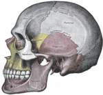
The human skull is a bony structure, part of the skeleton, that is in the human head and which supports the structures of the face and forms a cavity for the brain.
In humans, the adult skull is normally made up of 28 bones.[1] Except for the mandible, all of the bones of the skull are joined together by sutures, synarthrodial (immovable) joints formed by bony ossification, with Sharpey's fibres permitting some flexibility.
Components

Eight bones form the neurocranium (brain case), a protective vault of bone surrounding the brain and brain stem. Fourteen bones form the splanchnocranium, which comprises the bones supporting the face. Encased within the temporal bones are the six auditory ossicles of the middle ear. The hyoid bone, supporting the larynx, is usually not considered as part of the skull, as it is the only bone that does not articulate with other bones of the skull.
The skull also contains the sinus cavities, which are air-filled cavities lined with respiratory epithelium, which also lines the large airways. The exact functions of the sinuses are debatable; they contribute to lessening the weight of the skull with a minimal reduction in strength, they contribute to resonance of the voice, and assist in the warming and moistening of air drawn in through the nasal cavity.
Development of the skull

The skull is a complex structure; its bones are formed both by intramembranous and endochondral ossification. The skull roof, comprising the bones of the splanchnocranium (face) and the sides and roof of the neurocranium, are formed by intramembranous (or dermal) ossification, though the temporal bones are formed by endochondral ossification. The endocranium, the bones supporting the brain (the occipital, sphenoid, and ethmoid) are largely formed by endochondral ossification. Thus frontal and parietal bones are purely membranous.[2] The geometry of the cranial base and its fossas: anterior, middle and posterior changes rapidly, especially during the first trimester of pregnancy. The first trimester is crucial for development of skull defects.[3]
At birth, the human skull is made up of 44 separate bony elements. As growth occurs, many of these bony elements gradually fuse together into solid bone (for example, the frontal bone). The bones of the roof of the skull are initially separated by regions of dense connective tissue called "fontanels". There are six fontanels: one anterior (or frontal), one posterior (or occipital), two sphenoid (or anterolateral), and two mastoid (or posterolateral). At birth these regions are fibrous and moveable, necessary for birth and later growth. This growth can put a large amount of tension on the "obstetrical hinge", which is where the squamous and lateral parts of the occipital bone meet. A possible complication of this tension is rupture of the great cerebral vein of Galen. As growth and ossification progress, the connective tissue of the fontanelles is invaded and replaced by bone creating sutures. The five sutures are the two squamous, one coronal, one lambdoid, and one sagittal sutures. The posterior fontanel usually closes by eight weeks, but the anterior fontanel can remain open up to eighteen months. The anterior fontanel is located at the junction of the frontal and parietal bones; it is a "soft spot" on a baby's forehead. Careful observation will show that you can count a baby's heart rate by observing his or her pulse pulsing softly through the anterior fontanel.NNNNNNNNNNNNNNNNNNNNNNNNNNNNNNNNNNNNNNNNNNNNNNNNAAAAAAAAAAAAAAAAAAAAAAAAAAAAAAAAAAAAAAAAAAAAAAACCCCCCCCCCCCCCCCCCCCCCCCCCCCCCCCCCHHHHHHHHHHHHHHHHHHHHHHHHHHHHHHHHHHHHHHHHHHHHHHOOOOOOOOOOOOOOOOOOOOOOOOOOOOOOOOOOOOOSSSSSSSSSSSSSSSSSSSSSSSSSSSSSSSSSSSSSSSS ROCK!
Pathology
If the brain is bruised or injured it can be life-threatening. Normally the skull protects the brain from damage through its hard unyieldingness; the skull is one of the least deformable substances found in nature with it needing the force of about 1 ton to reduce the diameter of the skull by 1 cm.[4] In some cases, however, of head injury, there can be raised intracranial pressure through mechanisms such as a subdural haematoma. In these cases the raised intracranial pressure can cause herniation of the brain out of the foramen magnum ("coning") because there is no space for the brain to expand; this can result in significant brain damage or death unless an urgent operation is performed to relieve the pressure. This is why patients with concussion must be watched extremely carefully.
Dating back to Neolithic times, a skull operation called trepanation was sometimes performed. This involved drilling holes in the cranium. Examination of skulls from this period reveals that the "patients" sometimes survived for many years afterward. It seems likely that trepanation was performed for ritualistic or religious reasons and not only as an attempted life-saving technique. BONES: skull bones are divided into two main parts neurocranium(8) fascial skeleton(14)
Craniometry and morphology of human skulls
Like the face of a living individual, a human skull and teeth can also tell, to a certain degree, the life history and origin of its owner. Forensic scientists and archaeologists use metric and nonmetric traits to estimate what the bearer of the skull looked like. When a significant amount of bones are found, such as at Spitalfields in the UK and Jōmon shell mounds in Japan, osteologists can use traits, such as the proportions of length, height and width, to know the relationships of the population of the study with other living or extinct populations.
The German physician Franz Joseph Gall in around 1800 formulated the theory of phrenology, which attempted to show that specific features of the skull are associated with certain personality traits or intellectual capabilities of its owner. This theory is now considered to be obsolete. skull is basically a very important structure of human body and this leads to a very sensitivity to this tissue.
Sexual dimorphism
While in early life there is little difference between male and female skulls, in adulthood male skulls tend to be larger and more robust than female skulls which have boobs, which are lighter and smaller, with a cranial capacity about 10 percent less than that of the male.[5] However, the male body is larger than the female body, which accounts for the larger size of the male skull; proportionally, the male skull is about the same size as the female skull. Male skulls typically have more prominent supraorbital ridges, a more prominent glabella, and more prominent temporal lines. Female skulls generally have rounder orbits, and narrower jaws. Male skulls on average have larger, broader palates, squarer orbits, larger mastoid processes, larger sinuses, and larger occipital condyles than those of females. Male mandibles typically have squarer chins and thicker, rougher muscle attachments than female mandibles.

All of these features vary considerably within human populations, making it difficult to identify the sex of a skull without knowledge of the population from which it came.
Ancestry
Among archaeologists and forensic scientists, it is stated that the most consistent and unique trait of ancestry in a skeleton is its skull shape (see craniometry).[citation needed]
Additional images
-

-

-

-

-

-
-
 An cross-section of a skull by Leonardo da Vinci
 -
 Caucasian male human skull


Trivia
Unicode reserves character U+1F480 (💀) for a human skull pictogram.
See also
- Skull, a general article on skulls other than the human skull
- Base of the skull, detailed list of the anatomical structures found at the base of the skull
- Craniometry
- Foramina of the skull, list of holes (foramina) in the base of the skull
- Head and neck anatomy
- Obstetrical Dilemma
- Phrenology, the pseudoscientific process of determining personality from the shape of the head
- Plagiocephaly, the abnormal flattening of one side of the skull
- Human skull symbolism
- Anatomical terms of location
References
- ^ Skull Basics
- ^ Carlson, Bruce M. (1999). Human Embryology & Developmental Biology. Mosby. pp. 166–170. ISBN 0-8151-1458-3.
- ^ Derkowski, W; Kedzia, A; Glonek, M (2003). "Clinical anatomy of the human anterior cranial fossa during the prenatal period". Folia morphologica. 62 (3): 271–3. PMID 14507064.
- ^ Holbourn, A. H. S. (1943). MECHANICS OF HEAD INJURIES. The Lancet, 242: (6267), 438-441. doi:10.1016/S0140-6736(00)87453-X
- ^ "The Interior of the Skull". Gray's Anatomy. Retrieved 2010-111-28.
{{cite web}}: Check date values in:|accessdate=(help)