Gluteal tuberosity
| Gluteal tuberosity | |
|---|---|
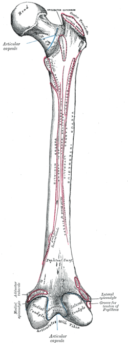
 Right femur. Posterior surface. (Gluteal tuberosity not labeled, but visible at upper right, where attachment of gluteus maximus is indicated.) | |
| Details | |
| Identifiers | |
| Latin | tuberositas glutaea femoris |
| TA98 | A02.5.04.017 |
| TA2 | 1376 |
| FMA | 43727 |
| Anatomical terminology | |
The gluteal tuberosity is the lateral one of the three upward prolongations of the linea aspera of the femur, extending to the base of the greater trochanter. It serves as the principal insertion site for the gluteus maximus muscle.
Structure
[edit]The gluteal tuberosity is the lateral prolongation of three prolongations of the linea aspera that extending superior-ward from the superior extremity of the linea aspera[1] on the posterior surface of the femur.[2]
The gluteal tuberosity takes the form of either an elongated depression[3] or a rough ridge. It extends from the linea aspera nearly vertically superior-ward to the base of the greater trochanter. Its superior part is often elongated to form a roughened crest, upon which a more or less prominent rounded tubercle - the third trochanter - is occasionally developed.[1]
Attachments
[edit]The gluteal tuberosity is the principal site of insertion of the gluteus maximus muscle,[4][5] accepting the muscle's tendon[6] (the gluteus maximus muscle additionally also inserts onto the iliotibial tract[5]).
References
[edit]- ^ a b Gray, Henry (1918). Gray's Anatomy (20th ed.). p. 246.
- ^ White, Tim D.; Folkens, Pieter A. (2005-01-01), White, Tim D.; Folkens, Pieter A. (eds.), "Chapter 15 - LEG: FEMUR, PATELLA, TIBIA, & FIBULA", The Human Bone Manual, San Diego: Academic Press, pp. 255–286, doi:10.1016/b978-0-12-088467-4.50018-1, ISBN 978-0-12-088467-4, retrieved 2021-02-27
- ^ Gray's anatomy : the anatomical basis of clinical practice. Susan Standring (Forty-second ed.). [New York]. 2021. p. 1362. ISBN 978-0-7020-7707-4. OCLC 1201341621.
{{cite book}}: CS1 maint: location missing publisher (link) CS1 maint: others (link) - ^ Chaitow, Leon; DeLany, Judith (2011-01-01), Chaitow, Leon; DeLany, Judith (eds.), "Chapter 11 - The pelvis", Clinical Application of Neuromuscular Techniques, Volume 2 (Second Edition), Oxford: Churchill Livingstone, pp. 299–389, doi:10.1016/b978-0-443-06815-7.00011-5, ISBN 978-0-443-06815-7, retrieved 2021-02-27
- ^ a b Waldman, Steven D. (2014-01-01), Waldman, Steven D. (ed.), "Chapter 84 - Gluteus Maximus Pain Syndrome", Atlas of Uncommon Pain Syndromes (Third Edition), Philadelphia: W.B. Saunders, pp. 244–247, doi:10.1016/b978-1-4557-0999-1.00084-8, ISBN 978-1-4557-0999-1, retrieved 2021-02-27
- ^ Gray's anatomy : the anatomical basis of clinical practice. Susan Standring (Forty-second ed.). [New York]. 2021. p. 1362. ISBN 978-0-7020-7707-4. OCLC 1201341621.
{{cite book}}: CS1 maint: location missing publisher (link) CS1 maint: others (link)
