
File:Oxy-heme Complex in Hemoglobin.png
Size of this preview: 800 × 520 pixels. Other resolutions: 320 × 208 pixels | 640 × 416 pixels | 846 × 550 pixels.
Original file (846 × 550 pixels, file size: 121 KB, MIME type: image/png)
| This is a file from the Wikimedia Commons. Information from its description page there is shown below. Commons is a freely licensed media file repository. You can help. |
Summary
| Description |
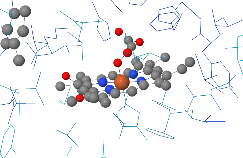
English: This figure was created using the Protein Data Bank and illustrates one of the four oxy-heme complexes with the coordinated O2 ligand |
| Date | |
| Source |
I created this image using The Protein Data Bank (https://www.rcsb.org/) which is a Public Domain archive, as per the following PDB Bank Usage Policies (https://www.rcsb.org/pages/usage-policy): "Images created using PDB data and other software should cite the PDB ID, the corresponding structure publication, and the molecular graphics program." "The wwPDB policy states that data files contained in the PDB archive are available under the CC0 1.0 Universal (CC0 1.0) Public Domain Dedication." |
| Author |
PDB Code 2DN1 1.25 a resolution crystal structures of human haemoglobin in the oxy, deoxy and carbonmonoxy forms. Park, S.-Y., Yokoyama, T., Shibayama, N., Shiro, Y., Tame, J.R. (2006) J Mol Biol 360: 690-701 DOI: 10.1016/j.jmb.2006.05.036 Image created using JSmol (Javascript) |
Licensing
| This file is made available under the Creative Commons CC0 1.0 Universal Public Domain Dedication. | |
| The person who associated a work with this deed has dedicated the work to the public domain by waiving all of their rights to the work worldwide under copyright law, including all related and neighboring rights, to the extent allowed by law. You can copy, modify, distribute and perform the work, even for commercial purposes, all without asking permission.
|
File history
Click on a date/time to view the file as it appeared at that time.
| Date/Time | Thumbnail | Dimensions | User | Comment | |
|---|---|---|---|---|---|
| current | 18:51, 24 September 2021 | | 846 × 550 (121 KB) | BillNye08 | Uploaded a work by PDB Code 2DN1 1.25 a resolution crystal structures of human haemoglobin in the oxy, deoxy and carbonmonoxy forms. Park, S.-Y., Yokoyama, T., Shibayama, N., Shiro, Y., Tame, J.R. (2006) J Mol Biol 360: 690-701 DOI: 10.1016/j.jmb.2006.05.036 Image created using JSmol (Javascript) from I created this image using The Protein Data Bank (https://www.rcsb.org/) which is a Public Domain archive, as per the following PDB Bank Usage Policies (https://www.rcsb.org/pages/usage-pol... |
File usage
The following pages on the English Wikipedia use this file (pages on other projects are not listed):