
Elbow: Difference between revisions
No edit summary |
|||
| Line 21: | Line 21: | ||
DorlandsSuf = 12161185 | |
DorlandsSuf = 12161185 | |
||
}} |
}} |
||
The '''elbow''' is the region surrounding the elbow-joint<ref>[http://www.emedicine.com/asp/dictionary.asp?keyword=Elbow eMedicine/Stedman Medical Dictionary Lookup!<!-- Bot generated title -->]</ref>—the ''ginglymus'' or [[hinge joint]] in the middle of the [[arm]]. Three bones form the elbow joint: the [[humerus]] of the upper arm, and the paired [[radius (bone)|radius]] and [[ulna]] of the forearm.<ref>[http://www.emedicine.com/asp/dictionary.asp?keyword=Elbow+joint eMedicine/Stedman Medical Dictionary Lookup!<!-- Bot generated title -->]</ref> |
The '''elbow''' is the knee of the arm. Its pit smells too. This region surrounding the elbow-joint<ref>[http://www.emedicine.com/asp/dictionary.asp?keyword=Elbow eMedicine/Stedman Medical Dictionary Lookup!<!-- Bot generated title -->]</ref>—the ''ginglymus'' or [[hinge joint]] in the middle of the [[arm]]. Three bones form the elbow joint: the [[humerus]] of the upper arm, and the paired [[radius (bone)|radius]] and [[ulna]] of the forearm.<ref>[http://www.emedicine.com/asp/dictionary.asp?keyword=Elbow+joint eMedicine/Stedman Medical Dictionary Lookup!<!-- Bot generated title -->]</ref> |
||
The bony prominence at the very tip of the elbow is the [[olecranon]] process of the ulna, and the inner aspect of the elbow is called the [[antecubital fossa]]. |
The bony prominence at the very tip of the elbow is the [[olecranon]] process of the ulna, and the inner aspect of the elbow is called the [[antecubital fossa]]. |
||
Revision as of 23:36, 25 June 2009
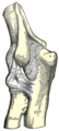
| Elbow-joint | |
|---|---|
 Elbow | |
 Left elbow-joint, showing anterior and ulnar collateral ligaments. | |
| Details | |
| Identifiers | |
| Latin | articulatio cubiti |
| MeSH | D004550 |
| TA98 | A01.1.00.023 |
| TA2 | 145 |
| FMA | 24901 |
| Anatomical terminology | |
The elbow is the knee of the arm. Its pit smells too. This region surrounding the elbow-joint[1]—the ginglymus or hinge joint in the middle of the arm. Three bones form the elbow joint: the humerus of the upper arm, and the paired radius and ulna of the forearm.[2]
The bony prominence at the very tip of the elbow is the olecranon process of the ulna, and the inner aspect of the elbow is called the antecubital fossa.
Movements
Two main movements are possible at the elbow:
- The hinge-like bending and straightening of the elbow (flexion and extension) ("joint") between the humerus and the ulna.
- The complex action of turning the forearm over (pronation or supination) happens at the articulation between the radius and the ulna (this movement also occurs at the wrist joint).
- The hinge moves in only one plane.
In the anatomical position (with the forearm supine), the radius and ulna lie parallel to each other. During pronation, the ulna remains fixed, and the radius rolls around it at both the wrist and the elbow joints. In the prone position, the radius and ulna appear crossed.
Most of the force through the elbow joint is transferred between the humerus and the ulna. Very little force is transmitted between the humerus and the radius. (By contrast, at the wrist joint, most of the force is transferred between the radius and the carpus, with the ulna taking very little part in the wrist joint).
Muscles, arteries, and nerves
The muscles in relation with the joint are:
- in front, the Brachialis, the Brachioradialis
- behind, the Triceps brachii and Anconæus
- laterally, the Supinator, and the common tendon of origin of the Extensor muscles
- medially, the common tendon of origin of the Flexor muscles, and the Flexor carpi ulnaris
The arteries supplying the joint are derived from the anastomosis between the profunda and the superior and inferior ulnar collateral branches of the brachial, with the anterior, posterior, and interosseous recurrent branches of the ulnar, and the recurrent branch of the radial. These vessels form a complete anastomotic network around the joint.
The nerves of the joint are a twig from the ulnar, as it passes between the medial condyle and the olecranon; a filament from the musculocutaneous, and two from the median.
Portions of joint
The elbow-joint comprises three different portions. All these articular surfaces are enveloped by a common synovial membrane, and the movements of the whole joint should be studied together.
| Joint | From | To | Description |
| humeroulnar joint | trochlear notch of the ulna | trochlea of humerus | Is a simple hinge-joint, and allows of movements of flexion and extension only. |
| humeroradial joint | head of the radius | capitulum of the humerus | Is an arthrodial joint. |
| proximal radioulnar joint | head of the radius | radial notch of the ulna | In any position of flexion or extension, the radius, carrying the hand with it, can be rotated in it. This movement includes pronation and supination. |
The combination of the movements of flexion and extension of the forearm with those of pronation and supination of the hand, which is ensured by the two being performed at the same joint, is essential to the accuracy of the various minute movements of the hand.
The hand is only directly articulated to the distal surface of the radius, and the ulnar notch on the lower end of the radius travels around the lower end of the ulna. The ulna is excluded from the wrist-joint by the articular disk.
Thus, rotation of the head of the radius around an axis passing through the center of the radial head of the humerus imparts circular movement to the hand through a very considerable arc.
Ligaments
The trochlea of the humerus is received into the semilunar notch of the ulna, and the capitulum of the humerus articulates with the fovea on the head of the radius. The articular surfaces are connected together by a capsule, which is thickened medially and laterally, and, to a less extent, in front and behind. These thickened portions are usually described as distinct ligaments.
The major ligaments are the ulnar collateral ligament, radial collateral ligament, and annular ligament.
Synovial membrane
The synovial membrane is very extensive. It extends from the margin of the articular surface of the humerus, and lines the coronoid, radial and olecranon fossæ on that bone; it is reflected over the deep surface of the capsule and forms a pouch between the radial notch, the deep surface of the annular ligament, and the circumference of the head of the radius. Projecting between the radius and ulna into the cavity is a crescentic fold of synovial membrane, suggesting the division of the joint into two; one the humeroradial, the other the humeroulnar.
Between the capsule and the synovial membrane are three masses of fat:
- the largest, over the olecranon fossa, is pressed into the fossa by the Triceps brachii during the flexion;
- the second, over the coronoid fossa,
- and the third, over the radial fossa, are pressed by the Brachialis into their respective fossæ during extension.
Terminology: "Elbow" , "Ell"
The now obsolete length unit ell relates closely to the elbow. This becomes especially visible when considering the Germanic origins of both words, Elle (ell, defined as the length of a male forearm from elbow to fingertips) and Ellbogen (elbow).
It is unknown when or why the second "l" was dropped from English usage of the word, but the elbow is shaped in an "L-shaped" unit, and thus the term elbow came to fruition.
Carrying angle

When the arm is extended, with the palm facing forward or up, the bones of the humerus and forearm are not perfectly aligned. The deviation from a straight line (generally on the order of 5-10°-men, 10-25°-women) occurs in the direction of the thumb, and is referred to as the carrying angle (visible in the right half of the picture, right). In females the carrying angle is greater than in males, due to the wider pelvic girdle exhibited in women.[3]
The carrying angle can influence how objects are held by individuals - those with a more extreme carrying angle may be more likely to pronate the forearm when holding objects in the hand to keep the elbow closer to the body.
Diseases of the elbow
The types of disease most commonly seen at the elbow are due to injury.
Tendonitis
Two of the most common injuries at the elbow are overuse injuries: tennis elbow and golfer's elbow. Golfer's elbow involves the tendon of the common flexor origin which originates at the medial epicondyle of the humerus (the "inside" of the elbow). Tennis elbow is the equivalent injury, but at the common extensor origin (the lateral epicondyle of the humerus).
Fractures
There are three bones at the elbow joint, and any combination of these bones may be involved in a fracture of the elbow. Patients who are able to fully extend their arm at the elbow are unlikely to have a fracture (98% certainty) and an X-ray is not required as long as an olecranon fracture is ruled out.[4]
Infection
Infection of the elbow joint (septic arthritis) is uncommon. It may occur spontaneously, but may also occur in relation to surgery or infection elsewhere in the body (for example, endocarditis).
Arthritis
Elbow arthritis is usually seen in individuals with rheumatoid arthritis or after fractures that involve the joint itself. When the damage to the joint is severe, fascial arthroplasty or elbow joint replacement may be considered.[5]
Additional images
-
 Medial Humerus Radius Ulna Articulated
 -
 Left Human Posterior Distal Humerus Extended
 -
 Left Human Posterior Distal Humerus Flexed
 -
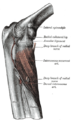
 Left elbow-joint, showing posterior and radial collateral ligaments.
 -
 Capsule of elbow-joint (distended). Anterior aspect.
 -
 Capsule of elbow-joint (distended). Posterior aspect.
 -
 The Supinator. Posterior view.
 -
 Diagram of the anastomosis around the elbow-joint.
 -
 Back of right upper extremity.
 -
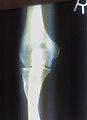 Close-up radiograph, right elbow-joint
 -
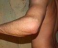 Left male elbow
 -
 Pathological fusion of three bones at elbow.


References
- ^ eMedicine/Stedman Medical Dictionary Lookup!
- ^ eMedicine/Stedman Medical Dictionary Lookup!
- ^ Steel, F (1958). "The carrying angle in man". Journal of Anatomy. 92 (2): 315–7. PMID 13525245.
{{cite journal}}: Unknown parameter|coauthors=ignored (|author=suggested) (help); Unknown parameter|day=ignored (help); Unknown parameter|month=ignored (help) - ^ Appelboam A; et al. (2008). "Elbow extension test to rule out elbow fracture: multicentre, prospective validation and observational study of diagnostic accuracy in adults and children". BMJ. 337: a2428. doi:10.1136/bmj.a2428.
{{cite journal}}: Explicit use of et al. in:|author=(help) - ^ Summary - Total elbow joint replacement for elbow arthritis: Surgery with a dependable, time-tested prosthesis can lessen pain and improve function in elbows, especially those with rheumatoid arthritis
![]() This article incorporates text in the public domain from page 321 of the 20th edition of Gray's Anatomy (1918)
This article incorporates text in the public domain from page 321 of the 20th edition of Gray's Anatomy (1918)