Deltoid muscle: Difference between revisions
m Reverted edits by 95.131.110.119 (talk) to last revision by Enfcer (HG) |
|||
| Line 31: | Line 31: | ||
===Origin and insertion=== |
===Origin and insertion=== |
||
It originates in three distinct sets of fibers:<ref>{{MedicalMnemonics|3558|||}}</ref> |
It originates in three distinct sets of fibers:<ref>{{MedicalMnemonics|3558|||}}</ref> |
||
*Anterior fibers: from |
*Anterior fibers: from |
||
*Lateral Fibers: from the lateral aspect of the [[acromion]] |
|||
*Posterior fibers: from the lower lip of the [[Posterior (anatomy)|posterior]] border of the [[spine of the scapula]], as far back as the triangular surface at its medial end.{{Citation needed|date=June 2011}} |
|||
From this extensive origin the fibers converge toward their insertion, the middle passing vertically, the anterior obliquely backward and laterally, the posterior obliquely forward and laterally; they unite in a thick [[tendon]], which is inserted into the V-shaped [[deltoid tuberosity]] on the middle of the [[Human anatomical terms#Anatomical directions|lateral]] aspect of the shaft of the [[humerus]]. At its insertion the muscle gives off an expansion to the [[deep fascia]] of the arm.{{Citation needed|date=June 2011}} |
|||
===Innervation and blood supply=== |
|||
The deltoid is innervated by the [[axillary nerve]].<ref name="Sunny">{{SUNYAnatomyLabs|03|03|01|03}}</ref> The axillary nerve originates from the ventral rami of the C5 and C6 cervical nerves, via the superior trunk, posterior division of the superior trunk, and the posterior cord of the brachial plexus.{{Citation needed|date=June 2011}} |
|||
The axillary nerve is sometimes damaged during operations on the [[axilla]], such as for [[breast cancer]]. It may also be injured by improper use of [[crutches]].{{Citation needed|date=June 2011}} |
The axillary nerve is sometimes damaged during operations on the [[axilla]], such as for [[breast cancer]]. It may also be injured by improper use of [[crutches]].{{Citation needed|date=June 2011}} |
||
Revision as of 15:04, 25 November 2011
| Deltoid muscle | |
|---|---|
 Deltoid muscle | |
 Muscles connecting the upper extremity to the vertebral column. | |
| Details | |
| Origin | the anterior border and upper surface of the lateral third of the clavicle, acromion, line of the scapula |
| Insertion | deltoid tuberosity of humerus |
| Artery | primarily posterior circumflex humeral artery |
| Nerve | Axillary nerve |
| Actions | shoulder abduction, flexion and extension |
| Antagonist | Latissimus dorsi |
| Identifiers | |
| Latin | musculus deltoideus |
| MeSH | D057645 |
| TA98 | A04.6.02.002 |
| TA2 | 2452 |
| FMA | 32521 |
| Anatomical terms of muscle | |
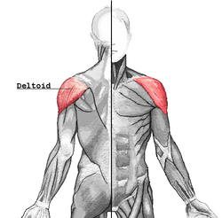
In human anatomy, the deltoid muscle is the muscle forming the rounded contour of the shoulder. Anatomically, it appears to be made up of three distinct sets of fibers though electromyography suggests that it consists of at least seven groups that can be independently coordinated by the central nervous system.[1]
It was previously called the Deltoideus and the name is still used by some anatomists. It is called so because it is in the shape of the Greek letter Delta (triangle). It is also known as the common shoulder muscle, particularly in lower animals (e.g., in domestic cats). Deltoid is also further shortened in slang as "delt". The plural forms of all three incarnations are deltoidei, deltoids and delts.[citation needed]
A study of 30 shoulders revealed an average weight of 191.9 grams (6.77 oz) (range 84 grams (3.0 oz)–366 grams (12.9 oz)) in humans.[2]
Human anatomy
Origin and insertion
It originates in three distinct sets of fibers:[3]
- Anterior fibers: from
The axillary nerve is sometimes damaged during operations on the axilla, such as for breast cancer. It may also be injured by improper use of crutches.[citation needed]
The deltoid is supplied by the posterior circumflex humeral artery.[4]
Action
When all its fibers contract simultaneously, the deltoid is the prime mover of arm abduction along the frontal plane. The arm must be medially rotated for the deltoid to have maximum effect[citation needed]. This makes the deltoid an antagonist muscle of the pectoralis major and latissimus dorsi during arm adduction.
The anterior fibers are involved in shoulder abduction when the shoulder is externally rotated. The anterior deltoid is weak in strict transverse flexion but assists the pectoralis major during shoulder transverse flexion / shoulder flexion (elbow slightly inferior to shoulders). The anterior deltoid also works in tandem with the subscapularis, pecs and lats to internally (medially) rotate the humerus.[5]
The posterior fibers are strongly involved in transverse extension particularly as the latissimus dorsi is very weak in strict transverse extension. Other transverse extensors, the infraspinatus and teres minor, also work in tandem with the posterior deltoid as external (lateral) rotators, antagonists to strong internal rotators like the pecs and lats. The posterior deltoid is also the primary shoulder hyperextensor, moreso than the long head of the triceps which also assists in this function.[6]
The lateral fibers perform basic shoulder abduction when the shoulder is internally rotated, and perform shoulder transverse abduction when the shoulder is externally rotated. They are not utilized significantly during strict transverse extension (shoulder internally rotated) such as in rowing movements, which use the posterior fibers.[7]
An important function of the deltoid in humans is stopping: preventing the dislocation of the humeral head when a person carries heavy loads. The function of abduction also means that it would help keep carried objects a safer distance away from the thighs to avoid hitting them, such as during a farmer's walk. It also ensures a precise and rapid movement of the glenohumeral joint needed for hand and arm manipulation.[2] The lateral fibers are in the most efficient position to perform this role, though like basic abduction movements (such as lateral raise) it is assisted by simultaneous co-contraction of anterior/posterior fibers.[citation needed]
In both the carrying of heavy loads and in lateral raises, the deltoid often contracts in tandem with scapular elevators such as the levator scapulae, upper trapezius or serratus anterior. By pulling the clavicle and scapulae up, it reduces compression and possibly impingement on the inferior borders so it doesn't press as much against the uppermost ribs.[citation needed]
The deltoid is responsible for elevating the arm in the scapular plane and its contraction in doing this also elevates the humeral head. To stop this compressing against the undersurface of the acromion the humeral head and injuring the supraspinatus tendon, there is a simultaneous contraction of some of the muscles of the rotator cuff: the infraspinatus and subscapularis primarily perform this role. In spite of this there may be still a 1–3 mm upward movement of the head of the humerus during the first 30° to 60° of arm elevation.[2]
Evolution
The deltoid is found in other apes. The human deltoid has a similar proportionate size to that of the muscles of the rotatory cuff to apes such as orangutans that engage in brachiation in which it holds the arm when used to the suspend the body. However in common chimpanzees the deltoid is much enlarged weighing 383.3g compared to the human average of 191.9g. In spite of this it is of less proportionate mass to the muscles of the chimpanzee rotatory cuff. This reflects the need in a knuckle walking ape to strengthen the shoulder, particularly the rotatory cuff, so it can support body weight.[2]
The deltoid is a classical example of a multipennate muscle.[citation needed]
The middle fibres of the muscle arise in a bipenniform manner (like a bird's feather) from the in number, which pass upward from the insertion of the muscle and alternate with the descending septa. The portions of the muscle arising from the clavicle and spine of the scapula are not arranged in this manner, but are inserted into the margins of the inferior tendon.[citation needed]
References
This article needs additional citations for verification. (June 2011) |
- ^ Brown JM, Wickham JB, McAndrew DJ, Huang XF. (2007). Muscles within muscles: Coordination of 19 muscle segments within three shoulder muscles during isometric motor tasks. J Electromyogr Kinesiol. 17(1):57-73. PMID 16458022 doi:10.1016/j.jelekin.2005.10.007
- ^ a b c d Potau JM, Bardina X, Ciurana N, Camprubí D. Pastor JF, de Paz F. Barbosa M. (2009). Quantitative Analysis of the Deltoid and Rotator Cuff Muscles in Humans and Great Apes. Int J Primatol 30:697–708. doi:10.1007/s10764-009-9368-8
- ^ MedicalMnemonics.com: 3558
- ^ Cite error: The named reference
Sunnywas invoked but never defined (see the help page). - ^ Template:Exrx
- ^ Template:Exrx
- ^ Template:Exrx
External links
- . GPnotebook https://www.gpnotebook.co.uk/simplepage.cfm?ID=-691011507.
{{cite web}}: Missing or empty|title=(help) - Template:MuscleLoyola
