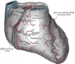
Coronary sulcus
| Coronary sulcus | |
|---|---|
 | |
 | |
| Details | |
| Identifiers | |
| Latin | sulcus coronarius |
| TA98 | A12.1.00.011 |
| TA2 | 3945 |
| FMA | 7174 |
| Anatomical terminology | |
The coronary sulcus (also called coronary groove, auriculoventricular groove, atrioventricular groove, AV groove) is a groove on the surface of the heart at the base of right auricle that separates the atria from the ventricles.[1][2] The structure contains the trunks of the nutrient vessels of the heart,[2] and is deficient in front, where it is crossed by the root of the pulmonary trunk. On the posterior surface of the heart, the coronary sulcus contains the coronary sinus.[3] The right coronary artery, circumflex branch of left coronary artery, and small cardiac vein all travel along parts of the coronary sulcus.
Structure
[edit]In relation to the rib cage, the coronary sulcus spans from the medial side of the 3rd left costal cartilage, to the middle of the right 6th costal cartilage.[1] Epicardial fat tends to be concentrated along the coronary sulcus.[4][5]
There are two coronary sulci in the heart, including left and right coronary sulci.
Left coronary sulcus
[edit]The left coronary sulcus originates posterior to the pulmonary trunk, and travels inferiorly separating the left atrium and left ventricle. The location of the left coronary sulcus is marked by the circumflex branch of left coronary artery, and coronary sinus.[6]
Right coronary sulcus
[edit]The right coronary sulcus begins anteriorly and superiorly on the sternocostal surface of the heart. Its position is marked by the location of the right coronary artery, and small cardiac vein. The right coronary sulcus separates the right atrium and its atrial appendage from the right ventricle inferiorly. The right coronary sulcus then passes inferiorly onto the diaphragmatic surface of the heart and traverses to the left.
Clinical significance
[edit]The left coronary sulcus is often neglected in echocardiography. As a result, normal variations and rare pathologic findings can be missed.[7]
See also
[edit]References
[edit]![]() This article incorporates text in the public domain from page 526 of the 20th edition of Gray's Anatomy (1918)
This article incorporates text in the public domain from page 526 of the 20th edition of Gray's Anatomy (1918)
- ^ a b Javadikasgari, Hoda; Gillinov, A. Marc; Mick, Stephanie; Mihaljevic, Tomislav; Suri, Rakesh M. (2019-01-01), Sellke, Frank W.; Ruel, Marc (eds.), "Chapter 21 - Robotic Mitral Valve Surgery", Atlas of Cardiac Surgical Techniques (Second Edition), Elsevier, pp. 347–363, doi:10.1016/b978-0-323-46294-5.00021-2, ISBN 978-0-323-46294-5, S2CID 81803257, retrieved 2020-11-16
- ^ a b Hargaden, Maureen; Singer, Laura (2012-01-01), Suckow, Mark A.; Stevens, Karla A.; Wilson, Ronald P. (eds.), "Chapter 20 - Anatomy, Physiology, and Behavior", The Laboratory Rabbit, Guinea Pig, Hamster, and Other Rodents, American College of Laboratory Animal Medicine, Boston: Academic Press, pp. 575–602, doi:10.1016/b978-0-12-380920-9.00020-1, ISBN 978-0-12-380920-9, retrieved 2020-11-16
- ^ Antonopoulos, Alexios S.; Siasos, Gerasimos; Antoniades, Charalambos; Tousoulis, Dimitris (2018-01-01), Tousoulis, Dimitris (ed.), "Chapter 2.1 - Functional Anatomy", Coronary Artery Disease, Academic Press, pp. 121–126, doi:10.1016/b978-0-12-811908-2.00008-8, ISBN 978-0-12-811908-2, retrieved 2020-11-16
- ^ Issa, Ziad F.; Miller, John M.; Zipes, Douglas P. (2019-01-01), Issa, Ziad F.; Miller, John M.; Zipes, Douglas P. (eds.), "27 - Epicardial Ventricular Tachycardia", Clinical Arrhythmology and Electrophysiology (Third Edition), Philadelphia: Elsevier, pp. 907–924, doi:10.1016/b978-0-323-52356-1.00027-x, ISBN 978-0-323-52356-1, retrieved 2020-11-16
- ^ Frühbeck, G.; Gómez-Ambrosi, J. (2013-01-01), "Adipose Tissue: Structure, Function and Metabolism", in Caballero, Benjamin (ed.), Encyclopedia of Human Nutrition (Third Edition), Waltham: Academic Press, pp. 1–13, doi:10.1016/b978-0-12-375083-9.00001-5, ISBN 978-0-12-384885-7, retrieved 2020-11-16
- ^ Ruberte, J.; Navarro, M.; Carretero, A.; König, H. E. (2017-01-01), Ruberte, Jesús; Carretero, Ana; Navarro, Marc (eds.), "11 - Circulatory System", Morphological Mouse Phenotyping, Academic Press, pp. 269–347, doi:10.1016/b978-0-12-812972-2.50011-8, ISBN 978-0-12-812805-3, retrieved 2020-11-16
- ^ Zuber, M.; Oechslin, E.; Jenni, R. (1998). "Echogenic structures in the left atrioventricular groove: diagnostic pitfalls". Journal of the American Society of Echocardiography. 11 (4): 381–386. doi:10.1016/S0894-7317(98)70107-5. ISSN 0894-7317. PMID 9571589.
External links
[edit]- Anatomy photo:20:st-1101 at the SUNY Downstate Medical Center
- Diagram
